
Life Science
INDUSTRY
Life Science
KEYWORD
INTRODUCTION SERVICE&PRODUCTS
Imaging mass spectrometry (IMS), which uses MALDI-TOF/MS analysis to identify the presence of target substances in biosamples such as tissue samples, is a cutting-edge measurement technology that is becoming increasingly popular in medical and biological research. The technology has potential not only for drug delivery and pharmacokinetics research, but also as a useful tool in biomarker research, and increasing numbers of measurements are now carried out using the iMScope TRIO. We sat down with Hidefumi Kaji, of Mitsubishi Tanabe Pharma Corporation's DMPK (Drug Metabolism and Pharmacokinetics) Research Laboratories, Research Division, and asked him about his use of this technology.

Hidefumi Kaji, Ph D
Group Manager
DMPK Research Laboratories, Research Division
*Affiliates and titles of the interviewee are current as of the time of reporting.
Mitsubishi Tanabe Pharma Corporation
URL
http://www.mt-pharma.co.jp/e/
You have been using the iMScope TRIO at the laboratory in Shimadzu's Sanjo Works for around two and a half years. What sort of research is it mainly used in?
I work in the DMPK Research Laboratories, and am engaged mainly in the creation of pharmaceutical products, specifically the area of preclinical DMPK. In my work, I aim to use the iMScope TRIO to examine the distribution of pharmaceutical candidate compounds in animal organs and tissue, and evaluate fluctuations in endogenous substances, biomarkers, and so on.
Could you please give us an actual example of pharmacokinetic analysis?
Of course. We used the iMScope TRIO to evaluate the distribution of a certain low molecular weight compound in rats. The compound we used is characterized by the fact that it bonds specifically to, and accumulate, the melanin in the eyeball. If a compound under development builds up by bonding to melanin or in some other way, we need to be careful about the photosensitivity of the compound. However, it's not always the case that if a compound bonds with melanin and is distributed within it, it demonstrates toxicity when it decomposes as a result of photosensitivity. Such understanding of what sort of degradation product occurs as a result of light is important in predicting the risk of toxicity. Furthermore, if we can establish that a compound dissipates relatively quickly, it usually means there will be no problems. From the pharmacokinetic standpoint, it is very important in the creation of safe drugs that we ascertain a compound's profile at an early stage of development, to see if it bonds to and accumulate melanin and what degradation products might occur.
I seem to recall that one of the main reasons you selected the iMScope TRIO was its spatial resolution.
That's right. The instrument is characterized by having a microscope at the front. The laser spec has improved significantly in the past few years, meaning that we can now perform high-resolution analysis, but I think that when analyzing a sample slice, the key point is being able to ascertain what point on the sample was being measured when the image in question was obtained. Having a microscope means that we can observe a part in detail and match results with what we are actually measuring. This has allowed us to correctly analyze at super-high resolution for the first time. Anyone who actually uses the instrument would be delighted by this. It is extremely important to position accurately and understand what part of the sample slice is being analyzed. The higher the resolution, the more likely it is that, just by moving five or ten microns, we are looking at a completely different place. The use of IMS has made it possible to confirm the specific distribution of the compound under evaluation within retinal melanin at super-high resolution, down to five microns. Many people have been amazed that we are able to examine samples in such detail. When people see the iMScope TRIO's high-resolution analysis in action for the first time, the impact is enormous.

Are there any other examples you can give us?
There is a substance called gentamycin, which is known to express toxicity within the kidneys in rats. There have been few reports that have proven where within the kidney it is distributed and expresses toxicity, and so we looked at whether it is possible to use IMS technology to evaluate this. Specifically, we implemented verification using 10-micron super-high-resolution analysis. Results showed that, when we looked at the distribution of the compound at a resolution that allowed the recognition of proximal tubules and glomerulus, gentamycin was distributed in proximal tubules, but had not built up in almost any other areas. In other words, we were able to confirm that the buildup of gentamycin in proximal tubules is the mechanism for toxicity expression.
Did you get more insightful analysis results thanks to using this technology?
Well, we're still at the basic research level, but we analyze biomarker fluctuation, which tells us what endogenous substances are active in a particular pathology caused by administration of a compound. For example, if gentamycin is administered and causes a disorder, it is assumed that the concentration of a particular endogenous substance will have risen compared with its state prior to administration. If we administer a drug or a compound that is a candidate for development, and the endogenous substance is reduced, allowing recovery from the disorder, the fluctuation in said endogenous substance indicates pathological improvement indirectly. Additionally, if the phenomenon is caused by the predicted efficacy mechanism of the development candidate compound, analyzing the phenomenon demonstrates that the drug has worked in line with its concept. Furthermore, if comprehensive analysis allows identification of types of endogenous substance, this means it may in the future allow the discovery of biomarkers that are clinically effective. This is an extremely attractive prospect.
So you are talking not only about drug discovery that aims for a cure, but also going a little further to the diagnostic stage. Early diagnosis and early cure are obviously important, but it is extremely interesting to hear how you are seeking biomarkers useful for determining the effectiveness of prognosis.
What are some of the conventional methods for evaluating tissue distribution?
In general, for a single project, a labeled compound is not synthesized at the point at which there are still multiple candidate compounds. Unlabeled compounds are administered, and organs subsequently removed, after which quantitative analysis is performed using LCMS on the homogenate of those organs.
And do you use autoradiography (ARG) after that, as part of the research process?
Yes. As the research stage progresses and we narrow down the compounds, we use radio-isotope (RI) labeled compounds to evaluate where a compound is distributed within the body after administration, where it builds up, and how long it takes before it is excreted, with the aim of applying for approval as a pharmaceutical product.
So you progress to ARG when you have narrowed down the candidate compounds?
That's right. In principle, we use it at the stage when a compound is moving into clinical testing. Creating markers requires significant investment, however, and we need to determine whether it is worth the investment cost. We try to avoid using ARG at the point where we still have multiple candidates.
What are the advantages and disadvantages of IMS in comparison with ARG?
The advantages are, firstly and most importantly, that we can perform evaluation at an early stage, prior to having to narrow the compounds down to one. The second relates to the greatest attribute of the mass spectrometer. With ARG, it is not possible to distinguish between the unaltered substance and metabolite distributions from marker tissue distribution information. In particular, when there are multiple metabolites present, the distribution and buildup information obtained is for a mixture of behaviors, where we don't know the ratio of the metabolites. Using a mass spectrometer allows us to see the distribution of each of the unaltered substances and metabolites. Members of the safety research section in our company have also said that they would like to use it to investigate toxicity mechanisms. This is because toxicity is sometimes caused by unaltered substances, and sometimes by metabolites.

Furthermore, it may be effective in the evaluation of safety considering the difference between animal species. One example is a case where some data shows a particular toxicity present in rats, which is caused by a metabolite that is only produced in rats, and therefore cannot be created in humans. Not only ARG but also mass spectrometry offers visual information. If it is possible to demonstrate that the toxicity is caused not by unaltered substance but rather by metabolites that are specific to rats, we can consider that the toxicity may not occur in humans.
That's very interesting!
As I just said, the great advantage is that it can be used not only for compounds but also for evaluating biomarkers that are active at the same time.
The non-targeting approach? That's a particular feature of this method. How about any disadvantages?
There is no guarantee that all compounds being measured can be ionized. That's the main disadvantage. In contrast to marker analysis, mass spectrometry can't detect non-ionized compounds, so it's not possible to use it to evaluate absolutely any and every compound.
That's true. That's the reason whole-body ARG probably won't be replaced completely with IMS, isn't it? How about sensitivity?
This is a very general comment, but MALDI currently compares poorly with LCMS and LC-MS/MS.
We are pressing ahead with improvements to sensitivity in pretreatment, thanks to Mitsubishi Tanabe Pharma's cooperation.
Yes, and then we need to think about what IMS is targeting, how much we can expect from it, and what it will be used for. If the target compound is highly ionized, which allows evaluation of effective dosage, then IMS can be used in efficacy mechanism analysis. But if it's not, then looking at efficacy evaluation levels might be difficult. When evaluating toxicity mechanisms, however, the dose and distribution quantity of the candidate compounds is sufficient, making their levels detectable. If IMS can be used for that purpose, it will offer attractive evaluation methods.
The microscopic level resolution is also part of the comparison to ARG, isn't it?
Given the problem of ionization, it is difficult to imagine that IMS completely replaces ARG when considering the current quantitative ARG results. The iMScope TRIO performs best when it is used not to evaluate whole-body distribution, but rather when it is focused on a certain type of tissue. This is because it allows high-resolution analysis. I think it is more appropriate to use it to look in a focused way at kidney tissue, brain tissue or liver tissue, and to get an accurate idea of how compounds, metabolites and biomarkers are distributed and fluctuate within those settings.
I see. In other words, it's not a replacement for ARG, it's a technology with the potential to provide supplementary information that cannot be obtained using ARG, and thus further help progress research and development in drug discovery.
Exactly. It's not possible to do everything using IMS, but we can use it with sample slices, to roughly check whether or not the target substances are present. Once we know where they are, we can perform more detailed analysis using mass spectrometry.
Both methods have advantages don't they? And these advantages can be used, rather than merely making up for the disadvantages.
So, looking ahead, what are your hopes for this technology in the future?
They are mainly to do with quantitative analysis. Pretreatment is important for this. Particularly, how evenly you can apply the matrix with good reproducibility. An automatic matrix application device is required for this. I think that the vaporization-type iMLayer, actually, is a very good device. When I present my results at seminars and so on, researchers looking at improving sensitivity and quantitative evaluation are always very interested in the automation aspect of the instrument.
How about areas other than quantitative analysis?
Chasing after the development compounds themselves is my major task using IMS, but I'd like to talk about another area. I am currently responsible for preclinical DMPK, and use laboratory animals for efficacy evaluation. I'd like to use the technology more in these evaluations. If the disease mechanism is a new one, efficacy evaluation becomes difficult. I'd like to discover biomarkers that can be used in that evaluation. If those biomarkers are reflected in human blood parameters when the drug goes into clinical trials, they can be used as indicators, and as markers to monitor treatment effectiveness when the drug is actually administered.

So you are talking about using IMS not only at an earlier stage in drug discovery than ARG, but also after entering the clinical phase.
I remember that in one of your lectures you said that, for biomarker research, the fluctuation results for biomarkers in the clinical phase would lead to the possibility of finding the next target. I was very impressed when I heard that.
Finally, do you have any opinions or hopes regarding the analytical instruments industry as a whole?
Within my area of work, which is mainly preclinical DMPK, we handle a lot of samples that originate in clinical research, and there is an increasing tendency towards working with materials actually used for clinical research. Since pharmaceutical companies exist to promote the health of patients, I would like to see Shimadzu working on the development of instruments that do not simply perform analysis for the sake of analysis, but rather take a step further towards the bigger vision of health care, for example by cooperating with us on the development of methods and instruments for use in analyzing markers that predict disease.
We certainly hope that you will continue to collaborate with us on the development of analysis instruments that can evaluate treatment as a whole, not only for diagnosis but also to confirm prognosis. Thank you for your insights and your time today.
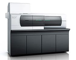
iMScope TRIO: Imaging Mass Microscope
We have had high expectations right from the start in regard to the uniqueness of the concept behind imaging mass spectrometry. Recently, not only have significant advances been made in the hardware, but significant progress has also been made in the field of pretreatment technology research aimed at practical utilization. My conversation with Mr. Kaji confirmed that IMS is being applied nowadays not only in basic medical research, but also in a variety of settings at the forefront of drug discovery research.
In response to this level of anticipation from our users, I feel strongly encouraged to continue with technological research that aims to "Realize Our Wishes for the Well-Being of Both Mankind and the Earth."