High Spatial Resolution Imaging by iMScope TRIO - Imaging of Chloroquine Distribution in Rat Retina -
Pharmacokinetic analysis of candidate compounds is a critical step in drug development, for elucidating the site and mechanism of activity and for evaluating local cytotoxicity.
Imaging mass spectrometry (MS) is a recently emerging technology as an alternative to conventional methods because it can quantify and give spatial distribution of both intact and metabolized forms of target compounds simultaneously. This label-free strategy is expected to drive drug development processes towards breakthrough discoveries. Here we present how imaging mass microscope iMScope TRIO was used for analysis of rat retina administered with chloroquine.
* Sales area: All areas excluding North America
High-Resolution Imaging of Chloroquine in Rat Retina
In this experiment, chloroquine, a well known drug against malaria infection, was administered to a rat to .elucidate its distribution in retina.
High resolution imaging of chloroquine distribution using a 10 μm section of pigment epithelium of retina was then performed by iMScope TRIO. The MS/MS product ion spectrum of chloroquine (m/z 320.190) and the plot of production intensity (m/z 247.095) were displayed as .Fig. 1 and 2, respectively.
Table 1 Analytical Conditions
| Sample Type and Preparation | |
| Sample | Chloroquine treated (20 mg/kg, po) rat retinal section (10 μm, OCT embedded) |
| MALDI Matrix | CHCA applied by sublimation |
| Analytical Conditions | |
| Polarity | positive mode |
| Precursor Ion | m/z 320.190 |
| Laser Diameter | 10 μm |
| Acquisition | 10 μm |
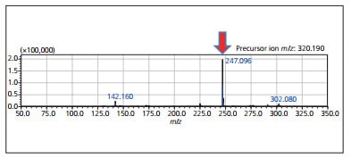
Fig. 1 MS/MS Product Ion Spectrum of Chloroquine Acquired Directly from Tissue Section
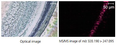
Fig. 2 MS/MS Imaging of Rat Retina, Showing the Spatial (Distribution of Chloroquine m/z 320.190 247.095)
iMScope TRIO
Imaging Mass Microscope
Imaging mass spectrometry is a revolutionary new technology.
The instrument is a combination of an optical microscope which allows the observation of high-resolution morphological images, with a mass spectrometer which identifies and visualizes the distribution of specific molecules.