SPM Observation of a Cross-Section
Introduction
Observation of the cross section of a specimen is often performed to further understand the properties of the specimen’s surface and to investigate the internal structure. Here, however, we present an example in which we used a scanning probe microscope (SPM) to observe the cross section of living tissue. As the specimen, we used an otolith taken from a bonito (migratory fish).
In general, pretreatment of a specimen is required before observing a cross section with a microscope. The pretreatment method varies with the purpose. In some cases, for example, the specimen is cut, embedded, and then cut again to reveal a cross section, and the specimen is observed in this state. In some cases, polishing or etching is performed before observation. Pretreatment is also required when using an SPM and so the pretreatment techniques are described and other relevant points are given.
Observation of a section of otolith increments in bonito
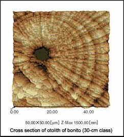
Fig.1 Image of Daily Rings
There are many migratory fish such as bonito and tuna that migrate from around the equator to the seas close to Japan. These species are referred to as “highly migratory”. Investigating the ecosystems of these types of fish is becoming an increasingly important activity in the context of monitoring and controlling resources trends. There are many points related to migratory habits and their development that are still unclear. Recently, researchers involved in the field of marine products have started trying to obtain greater knowledge by analyzing the daily rings on otoliths.
Using SEMs and optical microscopes, it has been deduced that the rings that appear on otoliths are daily rings. Investigating the pitch and number of these rings can give us a better idea about the mode of life of that species.
Here, we observed the daily rings for a bonito using an SPM, and confirmed that this is an effective instrument for this purpose. Fig. 1 shows the daily rings at the center of a cross section of an otolith that was taken from a 30-cm-class bonito. The ring shapes that resemble annual growth rings and the radial lines give us an idea about the growth of this fish.
Fig. 2 shows a profile image of the cross section. Conventional microscopes yield 2D images and can therefore be used only to ascertain the pitch and number of the rings. SPMs, however, yield 3D information and allow the topography to be seen in greater detail in profile display. This means that the changes that occur in one day of growth can be observed.
Fig. 3 shows a panorama image of fringes of the rings. Three sets of data have been combined to attain a wide area.
Pretreatment
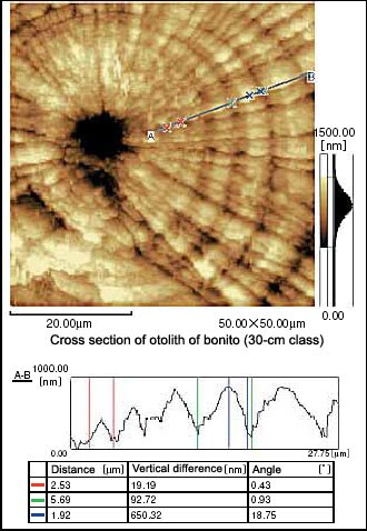
Fig.2 Profile analysis of daily rings
The high vertical resolution (0.1 nm) of the SPM is an important feature of this microscope. Special consideration is required, however, when observing cross sections. In comparison with other microscopes, the spatial resolution (in particular, the vertical resolution) is so good that scratches made during the cutting and polishing performed as part of pretreatment may be picked up. Therefore, to ensure that the structure under observation is not adversely affected, the cross section must be polished in order to make it flatter. Here, #6000-type wrapping tape was used to apply the last finish.
On the other hand, if polishing is performed perfectly and the cross section is rendered completely flat, because there will be no level differences in the structure of the cross section, it may not be possible to observe the desired structure. To counter this problem, the level differences within the structure are exposed using chemical etching, and the etching surface is observed using an AFM. Aqueous phosphoric acid solution (10%) is used as the etching fluid. Less etching is required than with other microscopes (1 s max.) and the level of structural damage resulting from excessive etching is minimized. After washing with water, the specimen is dried to complete pretreatment. The metal coating required with SEMs is unnecessary.
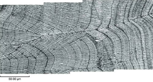
Fig.3 Panorama image of daily rings
This report was prepared with guidance from Mr. Tanabe of the Tohoku National Fisheries Research Institute, Fisheries Research Agency.