Brain Activity during Motor Control
In rehabilitation-related occupations, such as physical therapy, occupational therapy, and speech therapy, important aims include adaptive motor learning and recovery of motor function in patients. The ability of fNIRS to record brain activity during exercise and movement has resulted in its rapid adoption in the field of rehabilitation research.
The diagrams below show brain activity during basic limb movements (walking, upright posture control, arm reaching movements, and lower-limb muscle force control).
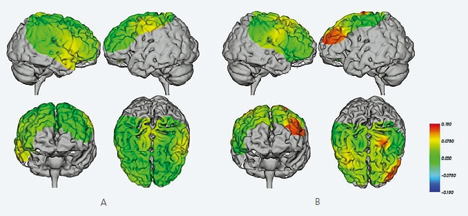
Brain Activity during Lower-Limb Locomotion (Walking on a Treadmill)
A: Brain images when walking at 4 km/hour. They confirm a slight increase in Oxy-Hb concentration-length (Oxy-Hb concentration multiplied by the path length) on both sides of the primary motor cortex compared to the resting state. They also show an increased Oxy-Hb concentration-length in the pre-motor cortex.
B: Brain images when avoiding obstacles at 4 km/hour walking speed. The Oxy-Hb concentration-length when walking at 4 km/hour is subtracted from the Oxy-Hb concentration-length while avoiding obstacles at 4 km/hour walking speed. The images confirm an increase in Oxy-Hb concentration-length in the right pre-motor cortex and left pre-frontal cortex (near the dorsolateral cortex).
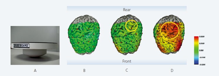
Brain Activity during Upright Posture Control
A: Wobble board used. B: Brain images when maintaining posture with feet together and eyes closed. The Oxy-Hb concentration-length in the reference state (maintaining upright posture with feet together and eyes open) is subtracted. C: Brain images when maintaining posture while standing on right leg. The reference state is subtracted. They confirm an increase in Oxy-Hb concentration-length in the right medial primary motor cortex and right pre-frontal cortex. D: Brain images when maintaining upright posture on a wobble board. They confirm an increase in Oxy-Hb concentration-length in the left and right primary motor cortex, supplementary motor cortex, and pre-frontal cortex.
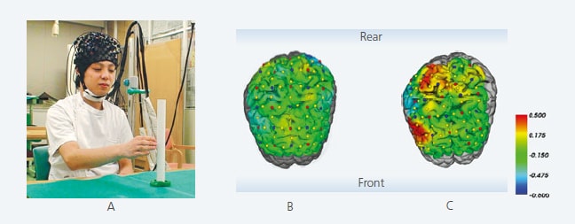
Brain Activity during Arm Reaching Movements
A: Photograph of test. B: No prism glasses. C: Wearing prism glasses. Cases of unilateral spatial neglect often occur where the patient is unable to concentrate on the left half of the visual field due to damage to the right hemisphere parietal lobe. A method known as prism adaptation is used for rehabilitation in such cases. This test was performed for the basic verification of the effects of the method during arm reaching movements. Image C confirms an increase in Oxy-Hb concentration-length in the right lateral pre-frontal cortex and parietal lobe in comparison with image B.
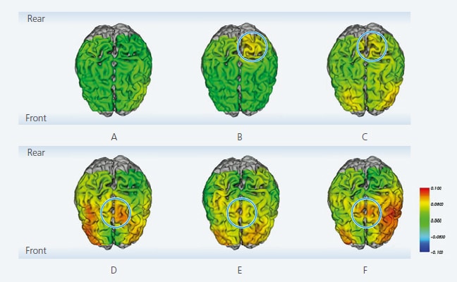
Brain Activity for Muscle Force Control during Right Knee Extension Movements
A: Brain image during Incremental exercise to 20 %MVC (maximal voluntary contraction). (The resting state is subtracted.) B: Incremental exercise to 40 %MVC. An increase in Oxy-Hb concentration-length is confirmed in the left medial primary sensorimotor cortex. C: Incremental exercise to 60 %MVC. An increase in Oxy-Hb concentration-length is confirmed in the left medial primary sensorimotor cortex and left and right pre-frontal cortex. D: Decreased exercise from 20 %MVC. E: Decreased exercise from 40 %MVC. F: Decreased exercise from 60 %MVC. Greater Oxy-Hb concentration-length is confirmed in comparison with incremental exercise in the left and right pre-frontal cortex and supplementary motor cortex.
(Data supplied by: Shu Morioka, MD, PhD, Department of Neurorehabilitation, Graduate School of Health Science, Kio University)
References:
Nobusako, S., Hiyamizu, M., Maeoka, H., Morioka, S.: Neurorehabilitation and Brain Function Imaging -- Walking (1), Physiotherapy 27: 274-282
Hiyamizu, M., Maeoka, H., Fujita, H., Morioka, S.: Neurorehabilitation and Brain Function Imaging – Upright Posture Control (2), Physiotherapy 27: 387-392, 2010 Taniguchi, H., Matsuo, A., Maeoka, H., Morioka, S.: Neurorehabilitation and Brain Function Imaging – Arm Reaching Movements (3), Physiotherapy 27: 499-504, 2010 Nobusako, S., Takebayashi, H., Hiyamizu, M., Maeoka, H., Morioka, S.: Neurorehabilitation and Brain Function Imaging – Muscle Force Control (5), Physiotherapy 27: 706-712, 2010
Note: The data shown was acquired using a FOIRE/OMM series model.
Related Products
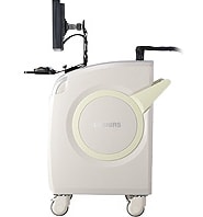
LABNIRS
functional Near-Infrared Spectroscopy System for Research
The laboratory model is ideal for a wide variety of basic research fields.
With a broad range of possible measurement regions, it can be readily customized for specific experimental conditions.
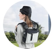
LIGHTNIRS
Portable functional Near-Infrared Spectroscopy System for Research
The portable model is ideal for field research.
It expands the possibilities for measuring brain function in a diverse range of applications and research fields.