Example of Brain-Function Imaging Research: Comparison of fNIRS and fMRI
Since fNIRS can measure not only Deoxy-Hb, but also changes in Oxy-Hb and blood flow, it is especially well suited to imaging brain function in patients where brain oxygen metabolite activity and hemodynamics are not normal. Currently, BOLD-fMRI is the primary method used to image brain function, but using it in conjunction with fNIRS allows accurately imaging brain function when the patient is sick.
A: Measurement by fMRI
Clearly shows activity in motor cortex for opposite side due to exercise (grasping). The lower BOLD* signal shows an increase during the task (blue).
B: Measurement by fNIRS
Displays results from 2D mapping (overlaid on MRI image) of Deoxy-Hb and Oxy-Hb during exercise. The lower graph shows the change in Oxy-Hb and Deoxy-Hb in the motor cortex (marked with circles). It shows how Oxy-Hb and Total-Hb increase and Deoxy-Hb decreases during the task (40 sec).
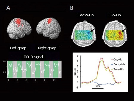
Comparison of BOLD-fMRI (A) and fNIRS (B) for Normal Healthy Adult
*BOLD: Blood Oxygen Level Dependent
Since fNIRS is not affected by electrical noise or magnetic fields and has few limitations on patient posture during measurements, it can be used to measure brain function in cases where BOLD-fMRI cannot be used, such as on patients being treated with deep brain stimulation (DBS) using a metal electrode.
Note: The data shown was acquired using a FOIRE/OMM series model.
Related Products
LABNIRS
functional Near-Infrared Spectroscopy System for Research
The laboratory model is ideal for a wide variety of basic research fields.
With a broad range of possible measurement regions, it can be readily customized for specific experimental conditions.

LIGHTNIRS
Portable functional Near-Infrared Spectroscopy System for Research
The portable model is ideal for field research.
It expands the possibilities for measuring brain function in a diverse range of applications and research fields.