


Article
Minimally Invasive Procedure in Practice ―Efforts by Ota Memorial Hospital―
Due to the increasing complexity and sophistication of interventional procedure in recent years, there is a demand for angiography systems capable of reducing exposure, reducing contrast media usage, and reducing examination times. Shimadzu's latest Trinias series of angiography systems is equipped with a variety of functions that facilitate minimally invasive treatment.
This article focuses on using fluoroscopy Low mode, radiography Low mode, and fluoroscopy record function to achieve additional reductions in radiation dose. The efforts of Ota Memorial Hospital (Gunma Prefecture, Japan) in using these functions to operate Trinias with low radiation doses are described.
1. Using Fluoroscopy Low Mode
During PCI and other interventional procedures, fluoroscopy accounts for a large proportion of the total treatment time that increases further when a complex lesion is being treated. For simple PCI, fluoroscopy is said to take approx. 10 times longer than radiography, and from the standpoint of dose management, reducing radiation dose of fluoroscopy is considered of prime importance.
Ota Memorial Hospital commonly uses a fluoroscopy program on Low mode with 7.5 pps frame rate (7.5 pps/Low) for the purpose of reducing the radiation dose of fluoroscopy.
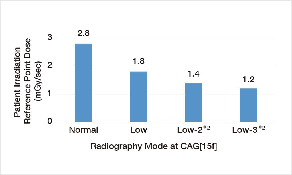
Fluoroscopy Low mode decreases the radiation dose by 44 % compared to fluoroscopy Normal mode (see figure), which allows for a substantial reduction in radiation dose over the course of a procedure. On previous systems, image lags were generated using modes with a frame rate of 7.5 pps, but Trinias uses SCORE PRO Advance proprietary image processing technology to reduce the appearance of Image lags. Ota Memorial Hospital has described Trinias with SCORE PRO Advance in fluoroscopy Low mode as "not an impediment to the procedure, and comparable in image quality to fluoroscopy Normal mode."

- Comparison of Radiation Doses by Fluoroscopy Mode
- Patient irradiation reference point dose with 8-inch FOV and 20 cm acrylic phantom.
(Measurement data that appears in Shimadzu's clinical manual. Low-2 and Low-3 values have been calculated.procedures)
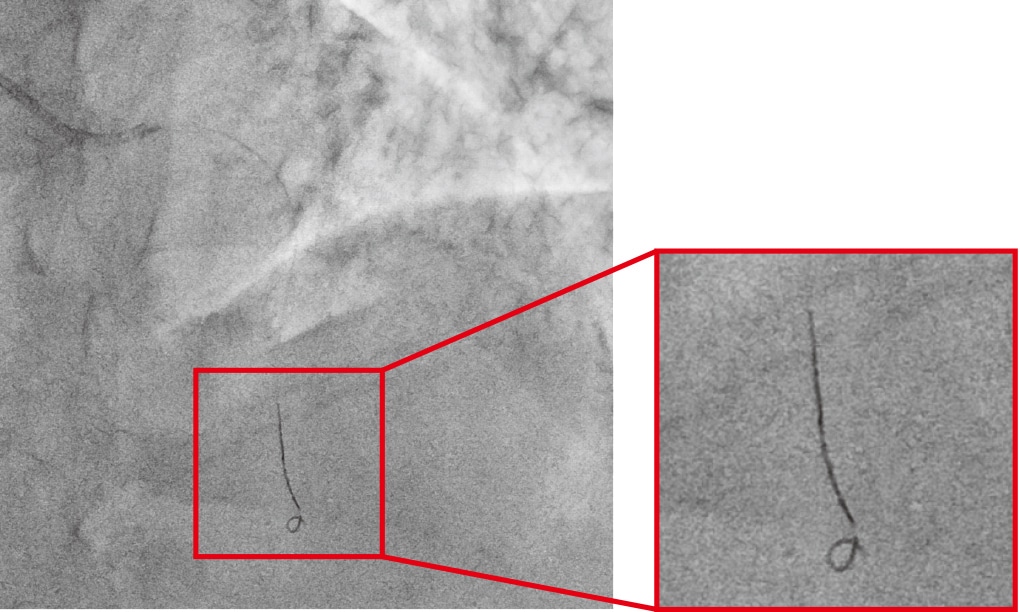
- Fluoroscopy Images Acquired at 7.5 pps/Low
- Guidewire is visible at low radiation dose and low frame rate without afterimages.

- A Word from Mr. Shigemitsu Hoshikawa, Department of Diagnostic Imaging
- A relatively large number of radiological technologists (13 people) work in rotation in the 2 catheterization rooms at Ota Memorial Hospital due to the conditions of the work that include night shifts and being on-call. Although this was the first time we procured a Shimadzu angiography system, we used SCORE StentView and SCORE RSM frequently from the beginning, and have found them to be effective for treatment and diagnosis. Introducing use of fluoroscopy Low mode to reduce exposure also occurred smoothly, and the system has been extremely easy to use. We intend to continue making use of these applications for the benefit of our work.

- A Word from Mr. Masahiro Fukasawa, Department of Diagnostic Imaging
- With advances in devices and other areas, an increasing number of treatments performed in cardiac and peripheral blood vessels now involve complex lesions. Ota Memorial Hospital has treated not a few cases of complex lesions. This lead to us giving serious consideration to reducing patient exposure, and to procuring Trinias in 2015 and after an investigation, introducing use of Low mode for both fluoroscopy and radiography. With SCORE PRO Advance, we obtain comparative image quality in Low mode as in Normal mode, and alongside using fluoroscopy record function we are performing treatment with low exposures.
Fluoroscopy Low mode is currently used by Ota Memorial Hospital regardless of the body type of the patient. System settings*1 are configured to allow instant switching to fluoroscopy Normal mode if needed, and Trinias is operated at all times to reduce radiation dose if possible, but ensure sufficient radiation dose if required by difficult circumstances. Trinias also has further settings for 2-steps of radiation dose reduction*2 that enable the radiation dose to be reduced by up to approx. 60% from fluoroscopy Normal mode, as required.
2. Using Radiography Low Mode
In PCI, image quality is extremely important for achieving better treatment outcomes. As one example, in the treatment of CTO lesions the presence of small collaterals (routes of collateral blood flow) has a major effect on treatment strategy, so it is important that images are of sufficient quality to show these structures. Since the radiation dose of radiography is also very high compared to fluoroscopy, reducing the radiation dose of radiography may also be effective in PCI.
At Ota Memorial Hospital, a Low mode radiography program (CAG[15f-15s]/Low) is regularly used to reduce the radiation dose of radiography. The radiation dose in radiography Low mode is reduced by 35 % from radiography Normal mode (see figure). Ota Memorial Hospital has described Trinias in radiography Low mode as "background granularity is increased somewhat, but there is adequate diagnostic performance for blood vessels." Trinias also has further settings for 2-steps of radiation dose reduction*2 for radiography programs that allows the user to reduce the radiation dose as required.
At Ota Memorial Hospital, discussion with doctors in the Department of Cardiology and technologists in the catheterization room have resulted in Low mode operation as the default setting for both fluoroscopy and radiography. At Ota Memorial Hospital, dose management is implemented through lively communication between doctors and technologists.
- Comparison of Radiation Dose by Radiography Mode
- Patient irradiation reference point dose with 8-inch FOV and 20 cm acrylic phantom.
(Measurement data that appears in Shimadzu's clinical manual. Low-2 and Low-3 values have been calculated.)
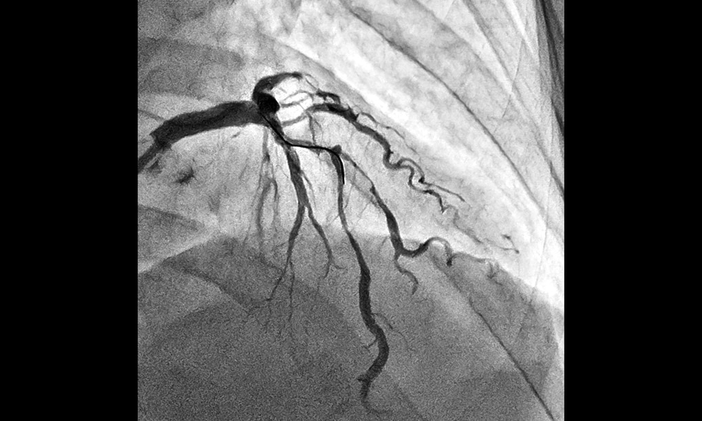
- Radiographic Images Acquired with CAG[15f-15s]/Low
- The background noise level is slightly higher than radiography Normal mode but the procedure is not impeded as major vessels are sufficiently visible.
3. Using Fluoroscopy Record Function
The status of balloon dilatation during PCI, lower extremity PPI, and other interventional procedures is retained in the form of images, though the method used to retain images of balloon dilatation differs by facility. The three methods used are (1) acquisition by radiography, (2) retain as a single image acquired with OneShot, and (3) use fluoroscopy record function to retain as a fluoroscopy image. The radiation dose of each method is highest for (1), followed by (2) then (3).
At Ota Memorial Hospital, after balloon dilatation has been observed by fluoroscopy, the state of dilation is recorded using fluoroscopy record function. Fluoroscopy image saving is operated with a single button and retains images instantaneously, thereby not impeding the progress of a procedure. Using fluoroscopy record function prevents increasing the radiation dose by the use radiography. Radiography is only performed for contrast imaging before treatment, to ascertain collateral location during treatment, and for confirmation contrast imaging after treatment, while fluoroscopy record function is used in all other circumstances when recording is required.
4. Dose Reduction Achieved by Efforts to Lower Radiation Dose
By implementing the above measures, Ota Memorial Hospital has reduced the radiation dose*3 for simple PCI by around 40 %.
Close coordination between doctors and technologists is valued at Ota Memorial Hospital, where both parties cooperated in carrying out a speedy investigation of Low mode usage.
- *1 Fluoroscopy Normal mode programs are allocated to function keys on a keyboard in the control room.
- *2 Configured by a service engineer on request.
- *3 Fluoroscopy record function was previously in use, so this effect is from use of fluoroscopy Low mode and radiography Low mode.
2018.3