Observation of plasmid DNA by SPM
DNA(deoxyribonucleic acid : which conveys genetic information in a living organisms) is the bio-polymer most studied by scanning probe microscopy methods. DNA was one of the first, if not the first biopolymer to be imaged by AFM (atomic force microscopy) and therefore there are many reports regarding this interesting target molecules(*1)
Figure 1 shows a three dimensional image of circular plasmid DNA molecules (plasmid vector (*2) , pGEM-Zf(+), purchased from Promega Inc., USA) deposited onto mica. This image was acquired in air with dynamic mode AFM using Shimadzu SPM-9500J2.
The procedure of the sample preparation is summarized in figure 2.
In the DNA imaging by AFM, sample needs to be deposited and secured onto a rigid substrate. The most commonly used substrate is mica. Since mica(natural mica : id called as muscovite, [KAI2(OH)2AISi3O10]) surface carries negative electrical charge, we are using divalent ions such as Mg to enhance the affinity between mica surface and the DNA molecules. Furthermore, mica should be freshly cleaved(split apart) just before the sample deposition to maintain very clean surface.
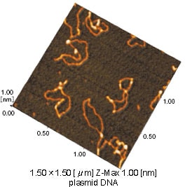
Fig.1 AFM image of plasmid DNA
This plasmid DNA is composed with about 3,000 base pairs of nucleotides. The contour length of this molecule (ca. 1 micro meter) calculated on this image is well suit with theoretical unit length (3.4 angstrom / base pair) which is well known by X-ray crystallography (Fig.3).
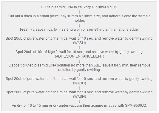
Fig.2 An example of the sample preparation procedure
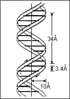
Fig.3 Molecular model of DNA
SPM has very high spatial resolution such as scanning electron microscope(SEM) and also it enables various kinds of sample observation in air, under solution and/or controlling the physiological conditions etc..
SPM imaging will provide :
a) ultra high resolution imaging by using carbon nano-tube tips,
b) analysis of molecular interaction between DNA and protein or other bio-molecules,
c) mapping of a sample surface depending of its chemical features,
and many other applications which could not be accomplished by any other imaging methods.
(*1)Referance
"Atomic Force Microscopy for Biologists", V.J.Morris, A.P. Gunning,
Imperial College Press, 1999
(*2)Special bio-molecule which is the genetic engineering to transport a gene into another cells is called as "vector", and a double stranded closed DNA named “plasmid” DNA is the most common vector for this purpose.