January 6, 2023 | News & Notices
High-Resolution*1 PET*2 System Provides Higher Image Quality than Conventional Systems
Expected to Contribute Accurate Diagnosis and Early Treatment of Dementia
Dr. Kazunari Ishii (Professor and Chairman, Department of Radiology, Kindai University Faculty of Medicine, Osaka-Sayama City, Osaka) and his research group demonstrated the diagnostic usefulness of using a world’s first high-resolution*1 PET*2 system capable of using for both head and breast imaging, a prototype of the TOF*3-PET System BresTome developed by Shimadzu Corporation (Kyoto) which can obtain higher quality (higher resolution) image than conventional whole-body PET systems. Accurate diagnosis is essential for early treatment of dementia. The TOF-PET system BresTome produces high-quality images that can provide benefit for the early treatment of dementia by enabling accurate diagnosis.
A paper on this work appeared online in the Journal of Nuclear Medicine (published by the Society of Nuclear Medicine and Molecular Imaging) on January 4, 2023.
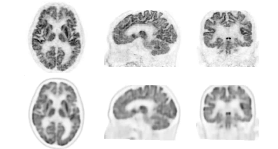
Top: Brain FDG-PET Images Obtained with TOF-PET System BresTome
Bottom: Brain FDG-PET Images Obtained with Conventional System
Top images appear of higher resolution and show greater detail. Bottom images appear somewhat blurry.
1. Key Points
- Images obtained with TOF-PET System BresTome are expected to enable more accurate diagnosis. The TOF-PET System BresTome, developed by Shimadzu Corporation, is the world’s first TOF-PET system that can be used for both head and breast imaging.
- Compared to conventional PET/CT systems, the TOF-PET System BresTome produces images of higher resolution that are expected to provide clinical benefits.
- Expected to benefit the early treatment of dementia by enabling early detection and accurate diagnosis. Early detection and accurate diagnosis are essential for the early treatment of dementia.
2. Background
Morphological imaging modalities such as CT and MRI measure physical structures, while PET is a functional imaging modality that measures metabolism and other physiological processes. When imaging the brain, PET can visualize processes such as glucose metabolism and the accumulation of substances that cause Alzheimer’s disease. However, compared to morphological imaging modalities, resolution of PET images is typically low and appear blurry. The development of PET system capable of high-resolution is highly desired.
3. Details
In October 2020, Kindai University Faculty of Medicine and Shimadzu Corporation began a clinical study at Kindai University Hospital using a prototype of the TOF-PET system BresTome developed by Shimadzu. The BresTome is a world’s first PET system capable of performing dedicated imaging of not only breasts but also head by improving previous dedicated breast PET system.
In the clinical study, PET images of glucose metabolism and amyloid*4 deposition primarily in patients with dementia are obtained with followed by BresTome and compared. Images obtained by BresTome showed excellent spatial resolution in all patients and even resulted in a change of diagnosis. These results suggested that BresTome contribute to accurate diagnosis in clinical practice and benefit the early treatment of dementia.
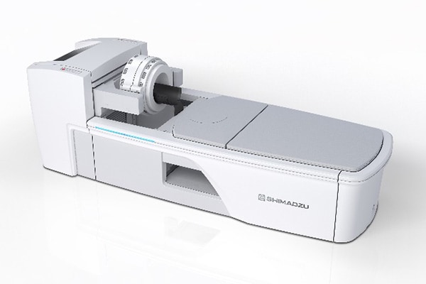
BresTome TOF-PET System (in Head Imaging Mode)
4. Published Paper
Publishing Journal: Journal of Nuclear Medicine (Impact Factor: 11.082 in 2021) Title: High-resolution silicon photomultiplier time-of-flight dedicated head PET system for clinical brain studies
Authors: Kazunari Ishii1,2*, Kohei Hanaoka2, Shota Watanabe2, Daisuke Ishikawa2, Takahiro Yamada2, Hayato Kaida1,2, Yoshiyuki Yamakawa3, Suzuka Minagawa3, Shiho Takenouchi3, Atsushi Ohtani3 and Tetsuro Mizuta3 * Corresponding author
Affiliations: 1. Department of Radiology, Kindai University Faculty of Medicine; 2. Division of Positron Emission Tomography, Institute of Advanced Clinical Medicine, Kindai University Hospital; 3. Medical Systems Division, Shimadzu Corporation
5. Details of Study
Shimadzu developed BresTome, the world’s first PET system that can be used for dedicated breast imaging and dedicated head imaging, by improving on the Elmammo dedicated breast PET system in current clinical use. The BresTome was developed with the goal of providing higher resolution images and higher sensitivity imaging than conventional PET/CT systems. To demonstrate clear imaging performance of BresTome against conventional PET/CT systems, specified clinical trial was performed at Kindai University Hospital that involved breast FDG-PET*5, brain FDG-PET*6, and brain amyloid PET*7 examinations. The study began with an investigation of dedicated head PET.
In the investigation, FDG images obtained by PET/CT and BresTome were compared for 18 patients with cognitive dysfunction, epilepsy, and other disorders. Next, amyloid images obtained by PET/CT and BresTome were compared for 17 patients with cognitive dysfunction. Images obtained by BresTome presented excellent resolution in all patients and greater resolution and visualized more detail than the PET/CT.
It has been noted that about 10% of brain amyloid PET scans are difficult to determine because the accumulation in gray matter is equivocal. In this investigation, one amyloid image obtained by PET/CT showed amyloid deposition in the right temporal lobe in all 17 patients. According to the imaging interpretation guidelines*8, this patient was diagnosed as positive for amyloid deposition (Alzheimer’s disease pathology is present). However, images obtained by BresTome showed no amyloid deposition in the right temporal lobe or any region of the brain. Based on this finding, the patient was judged negative for amyloid deposition and their diagnosis was changed.
Early detection and accurate diagnosis are essential for the early treatment of dementia. The findings of this clinical study are expected to contribute future clinical practice* at Kindai University Hospital.
* Note: Brain FDG-PET and brain amyloid PET examinations for dementia are not covered under national health insurance in Japan.
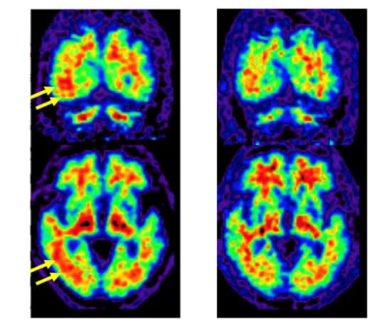
Left: Brain Amyloid PET Images Obtained with Conventional PET/CT System
Areas of PET tracer uptake indicated amyloid deposition. Yellow arrows show where PET tracer uptake appeared to extend to the gray matter of the brain (red areas extend to the surface of brain). Based on this, the gray matter was considered positive for amyloid accumulation.
Right: Brain Amyloid PET Images Obtained with BresTome
Images show that PET tracer uptake did not extend to gray matter (red areas do not extend to the surface of brain), which conflicts with the positive amyloid findings of the conventional PET/CT system.
6. Study Funding
This specified clinical trial (jRCTs052200055) received commissioned research funding from Shimadzu.
7. Glossary
*1. Resolution: The performance of a given system to delineate detail in an object
*2. PET: Acronym of positron emission tomography. PET is a functional imaging technique that involves administering a tracer labeled with a positron-emitting radionuclide, using a PET system to detect annihilation gamma-rays emitted by the radioactive tracer, and using this data to create images showing the degree of accumulation of the administered tracer.
*3. TOF (Time-of-flight): A technique used by PET systems that produces better image quality than non-TOF PET systems. The PET system determines the difference in detection time between two annihilation gamma rays and uses this difference to determine the position of the annihilated positron.
*4. Amyloid: Amyloid is a type of abnormal protein that is considered to cause Alzheimer’s disease by deposition and accumulation in the gray matter of the brain.
*5. Breast FDG-PET examination: Breast cancer shows high levels of glucose absorption. Breast FDG-PET examinations use this phenomenon to determine breast cancer malignancy based on degree of glucose uptake.
*6. Brain FDG-PET examination: Glucose is used by the brain as an energy source. Brain FDG-PET examinations use this phenomenon to show areas of reduced glucose consumption in the brain.
*7. Brain amyloid PET examination: Alzheimer’s disease is caused by amyloid proteins. Brain amyloid PET examinations use PET to show the presence or absence of amyloid deposits in the brain.
*8. Imaging interpretation guidelines: Imaging interpretation guidelines describe methods and standards for the interpretation of amyloid PET images and the identification of features that represent amyloid deposition (positive) or an absence of amyloid deposition (negative).