November 1, 2022 | News & Notices
With Three Types of Images, Contributing to Research and Development of New Materials
Release of the Xctal 5000, the First Phase-Contrast X-Ray CT System Produced in Japan

Product Photo: Xctal 5000 phase-contrast X-ray CT system
Shimadzu has released the Xctal 5000 phase-contrast X-ray CT system in Japan and other countries. This is the first phase-contrast X-ray CT system produced in Japan. It can create three types of images from a single scan: X-ray absorption images, dark-field images, and phase images. By using X-ray phase contrast, which images the scattering and the refraction of X-rays transmitted through materials, it can observe samples that are hard to observe with previous X-ray CT systems, which use the X-ray absorption contrast that images the absorption of X-rays. This new system will contribute to research and development of fiber reinforced resins, composite materials, and biomaterials.
X-ray CT systems irradiate samples rotating between an X-ray generator and detector with X-rays, and observe and scan the interior of the sample in three dimensions. The interior can be observed non-destructively, so these systems are used for both quality control and research and development. In recent years, R&D of new materials has accelerated with the trend toward carbon neutrality. The demand for non-destructive observations, such as inspections of the interior of CFRP, a lightweight material for automobiles, and observations of adhesively bonded joints in new materials combining different resins, is increasing. However, previous X-ray CT systems had difficulties providing both a large field of view and good resolution, and making high-contrast observations between materials with similar absorption coefficients.
This system adopts a new method (X-ray phase imaging technology) using a diffraction grating, enabling both dark-field image scans and phase image scans. Dark-field images can provide scans of tiny scratches inside samples, and the flow of fiber bundles over a wide field of view. They will likely be used to find more stable manufacturing methods for CFRP and other new materials. Phase images can provide high-contrast scans between materials with similar absorption coefficients, as long as the density difference is above a certain amount. They can also be used for observations of new resin materials and biological soft tissues.
Shimadzu Corporation has been working with X-ray instruments for more than a century and is a leading company in this field in Japan. We will continue to take the initiative in developing new products that provide new value.
Features
1. Both Wide Field of View and Detailed Structural Observations
The problem with conventional absorption images is that in order to observe detailed structure, high-magnification scans are required, which means narrowing the field of view and finely cutting the sample. This system uses dark-field images, so it can observe minute structures, including fine scratches, within a sample or the flow of fiber bundles. The system can scan a wide field of view, up to 100 mm per scan, enabling it to identify the presence of fine scratches over the entire sample and estimate their positions. There is no need to perform multiple scans or finely cut the sample.
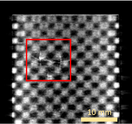
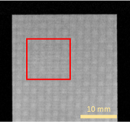
CFRP cross material with cracks
Cross-sectional image (Dark), Conventional cross-sectional image (Absorption)
2. Provides Two Types of High-Contrast Observations
In addition to absorption images, the Xctal 5000 can create phase images, enabling it to make high-contrast observations even between materials that have previously been hard to contrast. The range of observations is broadened by using two image types: absorption images, which visualize differences in absorption coefficients, and phase images, which visualize density differences.
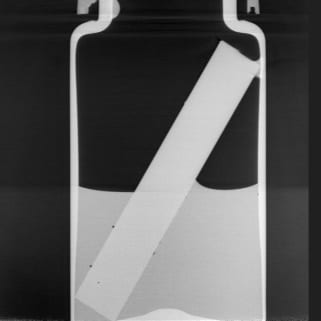
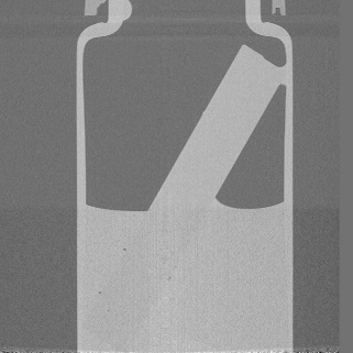
Water and Acrylic
Cross-sectional image (Phase), Conventional cross-sectional image (Absorption)
3. Anyone Can Scan Easily
To start scanning, just place the sample in the instrument and select preset scanning conditions. After scanning, observation and data analysis software suited to the scanning method starts up automatically, so data analysis is easy.
For more details, visit
Xctal 5000 phase-contrast X-ray CT system