November 4, 2020 | News & Notices
Shimadzu’s New Imaging System Enables Integrated Analysis of Both Trace Elements and Various Compounds
Advancing Elucidation of the Mechanisms of Disease Onset and the Development of Therapeutic Drugs
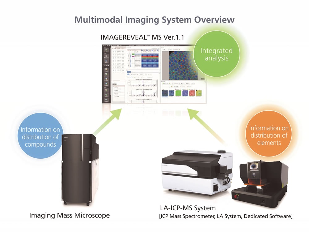
Shimadzu Corporation announces the release of a multimodal imaging system capable of separately acquiring information on the distribution of trace elements and the distribution of various compounds, and performing an integrated analysis of both sets of information. The system consists of a laser ablation inductively coupled plasma mass spectrometer (LA-ICP-MS) that acquires information on the distribution of elements, an imaging mass microscope that acquires information on the distribution of compounds, and the IMAGEREVEAL MS mass spectrometry imaging data analysis software that performs an integrated analysis of data acquired by the LA-ICP-MS and imaging mass microscope. The system is able to acquire more accurate information on the function of metals and metabolic mechanisms in the body.
With this multimodal imaging system, Shimadzu aims to advance the elucidation of mechanisms of disease onset and the development of therapeutic drugs in the life sciences. The technology found in the LA-ICP-MS is the result of collaborative research between Shimadzu’s European Innovation Center and Dr. Uwe Karst of the University of Münster.
Laser ablation (LA) involves irradiating a very small area of a solid sample with a laser to vaporize and generate particles from that part of the sample. Inductively coupled plasma mass spectrometry (ICP-MS) measures the types of elements and elemental content of a sample. The LA-ICP-MS combines these two technologies to enable investigation of trace element distributions in biological samples, mineral ore, semiconductors, metal, and other solid samples. The imaging mass microscope consists of an optical microscope combined with a MALDI mass spectrometer and is capable of visualizing the distribution of pharmacologically active constituents and various other compounds in biological samples.
Features of the Multimodal Imaging System
1. Integration of Complementary Imaging Technologies
The imaging mass microscope acquires information on the distribution of organic compounds and the LA-ICP-MS acquires information on the distribution of metals and other trace elements while also offering excellent quantitative performance. By combining these complementary technologies, advanced imaging analysis can be performed on a larger quantity of information.
2. Analysis of Analytical Samples at Atmospheric Pressure and Better Spatial Resolution Performance
Both the imaging mass microscope and the LA-ICP-MS analyze samples from a sample chamber that is at atmospheric pressure; hence, even biological samples containing water can be measured with ease. A spatial resolution performance of several μm to several tens of μm is also perfect for evaluating the distribution of target constituents in a very small area.
3. Integrated Data Analysis Software
The IMAGEREVEAL MS integrated data analysis software analyzes imaging data from the imaging mass microscope and the LA-ICP-MS from a variety of perspectives based on simple operating procedures that require minimal time and labor. IMAGEREVEAL MS reads and immediately analyzes data from both the imaging mass microscope and the LA-ICP-MS without the need for special data conversion.
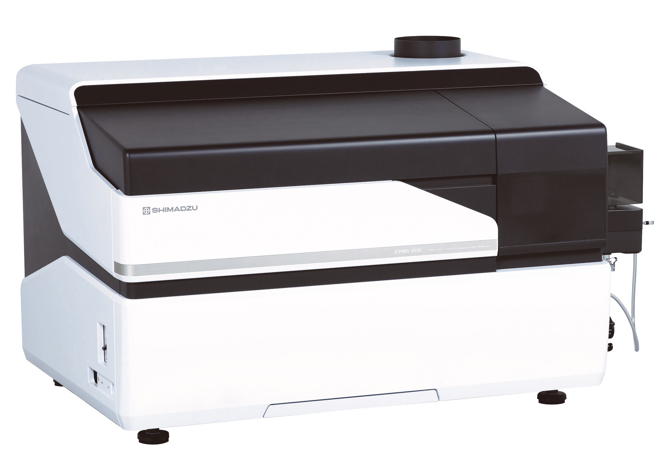
ICPMS-2030 Inductively Coupled Plasma Mass Spectrometer
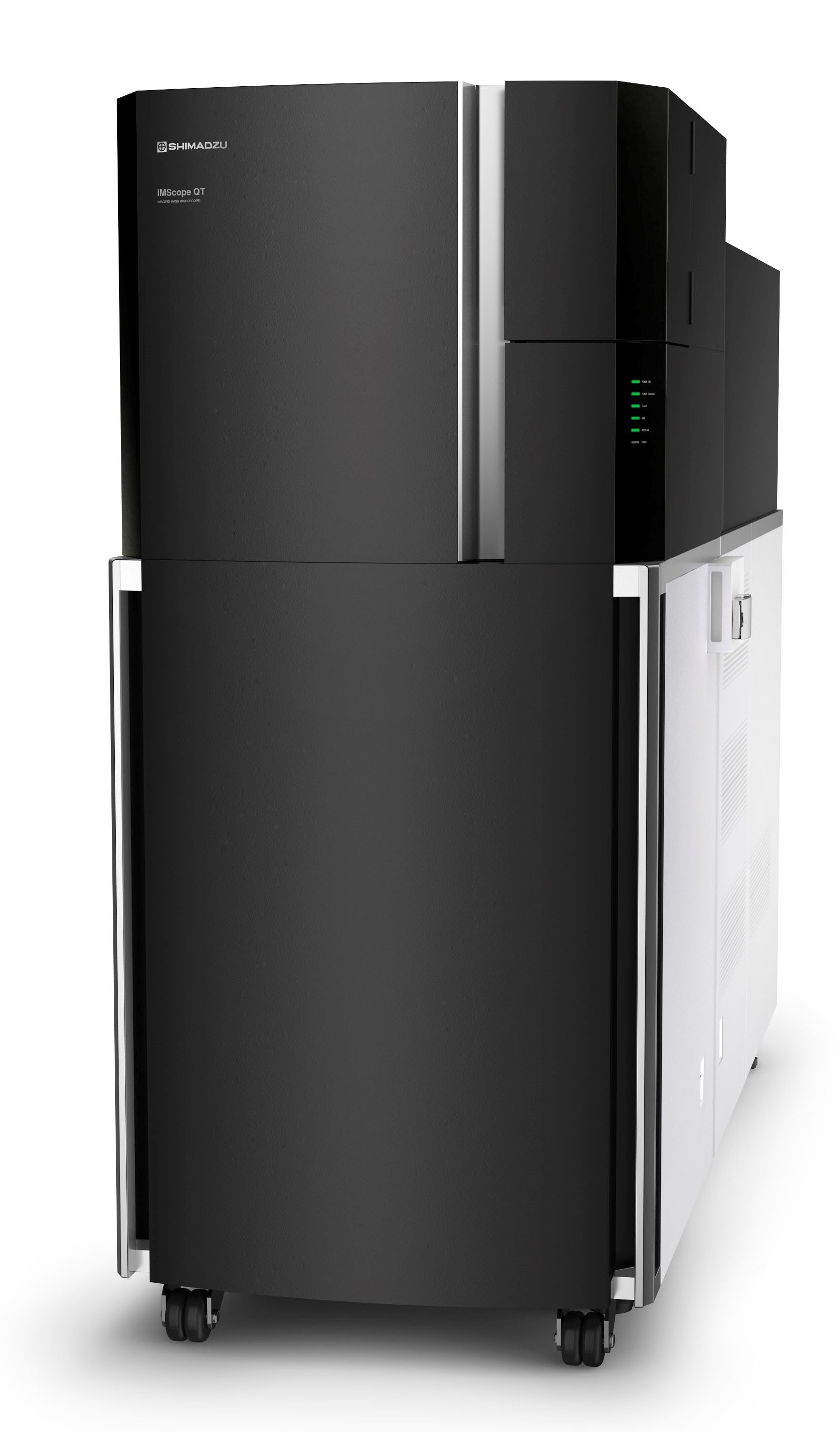
iMScope QT Imaging Mass Microscope
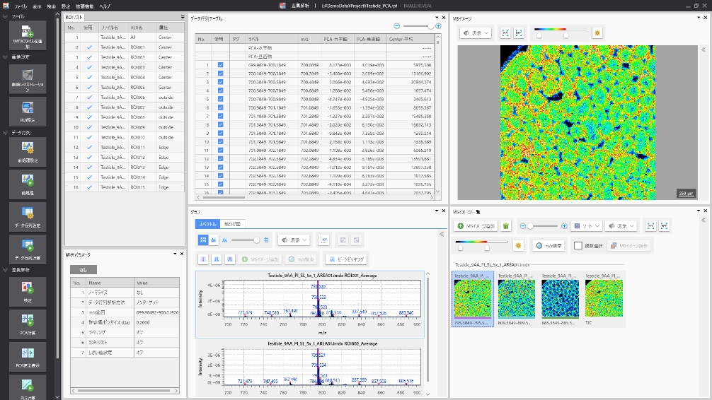
IMAGEREVEAL MS Mass Spectrometry Imaging Data Analysis Software
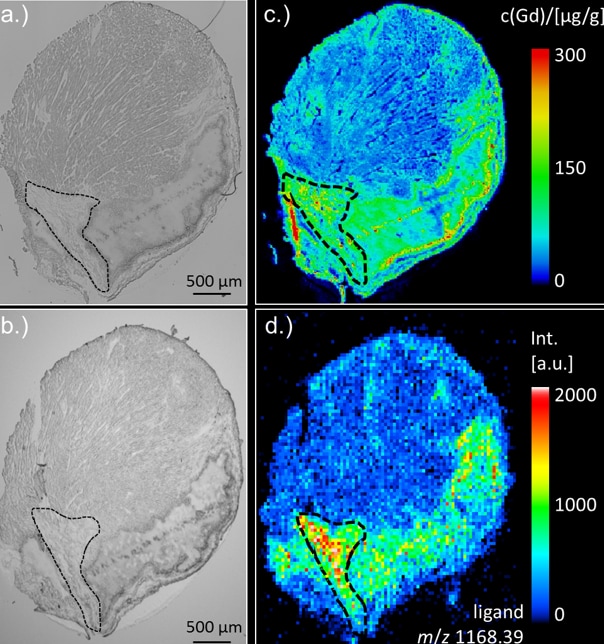
Example Analysis: Contrast Medium in Mouse Cardiac Tissue
- A gadolinium contrast medium exhibits a high affinity for the extracellular matrix secreted in cases of myocardial infarction. A gadolinium contrast medium was administered to a myocardial infarction model mouse and the distribution of the medium in mouse cardiac tissue was observed. The LA-ICP-MS was used to perform elemental imaging of the gadolinium contained in the contrast medium (c) and iMScope was used to perform molecular imaging of molecules with the same structure as the contrast medium (d). Combining these two sets of imaging data enables visualization of the distribution of gadolinium contrast medium in the cardiac tissue of the myocardial infarction model mouse. Images (a) and (b) were acquired by the optical microscope unit. The region affected by myocardial infarction is shown within the black dashed line.
For more details, visit
Multimodal Imaging System