Carbon Nanotube | Dispersion | Observations and Potential Measurements
The potential at and around carbon nanotubes (CNTs) can be measured on an electric force microscopy (EFM) image taken with a scanning probe microscope (SPM).
Fig. 1 shows a sample topographic image and an EFM image for an 8 wt% SWNT/acrylic resin sample.
Fig. 2 also shows an example of EFM measurements. The sample is a mixture of purified single-walled carbon nanotubes (HiPco) and DNA solution spin-coated onto a mica substrate. (Manufactured by Carbon Nanotechnology Inc.)
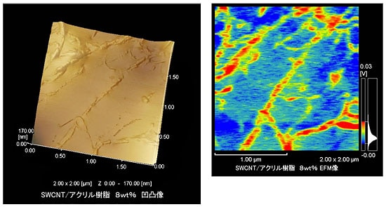
Fig. 1 Sample Topographic Image and EFM Image for 8 wt% SWNT/Acrylic Resin Sample (Samples supplied by Aida Nanospace Project, Japan Science and Technology Agency)
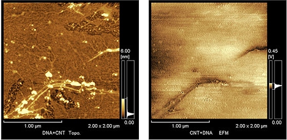
Fig. 2 EFM Measurements (Purified SWNTs (HiPco) and DNA Solution Spin-Coated onto a Mica Substrate) (Samples supplied by Shinohara Laboratory, Graduate School of Science, Nagoya University.
Scanning Probe Microscope
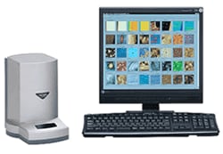
The scanning probe microscope (SPM) scans sample surfaces with a microscopic probe to provide high-magnification 3-dimensional observations.
The SPM-9600 is the next generation of spanning probe microscope that represents further evolution of previous models that were highly regarded for rapid observations and simple operation.