MS Imaging of Paraffin-Embedded Tissue Sections
MS imaging of paraffin-embedded tissue sections of human endometrial cancer and ovarian cancer
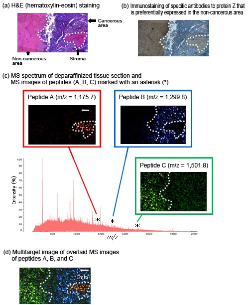
After de-paraffinization and enzyme treatment, MALDI-TOF MS imaging analysis is possible on paraffin-embedded tissue sections obtained from long-term storage at many hospitals and research organizations. Endometrial cancer sections (10 µm thick) stored for 7 years were de-paraffinized using hot xylene; trypsin and DHB matrix were dispensed with a CHIP-1000 Chemical Printer; and analysis performed by AXIMA-QIT.
Similarly, paraffin-embedded tissue sections of human ovarian cancer were de-paraffinized, trypsin and DHB matrix added by CHIP-1000, and MS/MS analysis performed using an AXIMA-QIT to directly identify the proteins in the sections. This allowed the detection and identification of multiple proteins, particularly the structural proteins comprising the cytoskeleton.
| Reference: | Yutaka Aoki et al (2007), "Potentiality of Drug Discovery with MALDI-MS Imaging System," Shimadzu Review 63 (3/4) |
MALDI-TOF MS Imaging System
- CHIP-1000 allows highly reproducible MALDI-TOF MS imaging.
- Allows the MALDI-TOF MS imaging, identification, and structural analysis of diverse molecules, ranging from lipids and low-molecular compounds to peptides and proteins.
- The acquired MS data can be read and analyzed using existing MS imaging software, such as BioMap (http://www.maldi-msi.org/).