Development of World’s First Remote-controlled X-ray R/F System for Patient and Healthcare Practitioner Safety
Series: Shimadzu's Heritage
In 1961, Shimadzu Corporation created the world’s first Remote-controlled X-ray R/F (Radiography and Fluoroscopy) System. An X-ray R/F system is an examination device that shows the inside of the body in real time by exposing it to X-rays.
The idea behind the development of this medical diagnostic device was to “minimize exposure of radiation not only to patients but also to the medical professions conducting the X-ray examination.” This product concept has been inherited as the basic philosophy for developing imaging diagnostic devices at Shimadzu Corporation. This article focuses on the story of how this technology was developed.
Lifestyle Diseases Changed the Situation
In the 1950s, Japan was in the midst of unprecedented rapid economic growth, and many people were enjoying a feeling of affluence. Improvements to public health and the emergence of new medicines helped to increase the lifespan and improve the nutrition of the Japanese people. However, the change from a traditional simple diet to one heavier in meats, fats, and oils, as well as changes to lifestyles, created new problems.
During this time, stroke, cancer, heart disease, and other “lifestyle diseases” were increasing yearly as causes of death for Japanese people. People in their working prime, from ages 40 to 60, were susceptible to these diseases, and the diseases were said to become more likely as they aged. An urgent need was seen at that time to establish a system for the early detection and treatment of these diseases. As a national policy, Japan began the introduction of medical checkups using a group screening system, whereby the members of a company, local public body, or other organization would get checked all at once as a group.
At the time, gastric cancer with the highest prevalence among Japanese people, and particular effort was put into screening for it. Patients were asked to drink a contrast medium to make the stomach easier to see, and examiners would then observe the actively moving stomach in real time to detect signs of cancer. In this way, X-ray examination devices were used to screen for gastric cancer.
Since Shimadzu launched sales of the first X-ray examination devices in Japan in the early 20th century, research into diagnosis and treatment technologies using X-ray technology has continued to make rapid advances. At the time, however, although these devices supported human health, they did not sufficiently protect the safety and health of the doctors, nurses, clinical X-ray technicians (now called “clinical radiologists”), and other practitioners administering X-ray examinations. This is because to observe moving organs, practitioners needed to keep operating the X-ray examination device by staying beside it.
Back then, to observe the stomach in real time, after a patient drank the contrast medium, practitioners had to irradiate a fluorescent plate with X-rays transmitted through the human body, and directly observe the image projected on the plate. Technicians were required to wear protective clothing to protect their body from the X-rays, but the protective clothing could not protect the thyroid gland or the crystalline lens from radiation, nor did it sufficiently reduce the total amount of radiation exposure. It was clear that cumulative exposure from repeated observations on a day-to-day basis would have harmful effects on the human body.
Another issue was the need for a darkroom. The light that emerged from the X-ray image was unreliable and could by no means be verified under illumination. After spending time to adjust their eyes to the dark, doctors and technicians would operate the device by touch. They had difficulty positioning patients for taking images, and would commonly search for lesions in the faint light of the images while being exposed to X-rays.
Despite the desire to increase the number of X-ray examinations for early diagnosis and treatment, a screening room could handle no more than about five gastric cancer screenings in a half-day period. A technological breakthrough was urgently needed to advance group screenings.
World’s First System Meets Wishes of Healthcare Practitioners
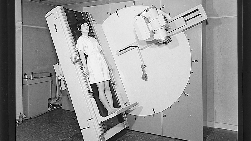
This situation was resolved in 1961 with the launch of the Remote-controlled X-ray R/F System. This was the world’s first system of its kind, developed by Shimadzu, together with Osaka Prefectural lifestyle diseases center (currently Osaka International Cancer Institute) and Matsushita Electric Industrial (currently Panasonic), at the request of the Osaka Prefectural government, which aimed to establish a new checkup system for gastric cancer.
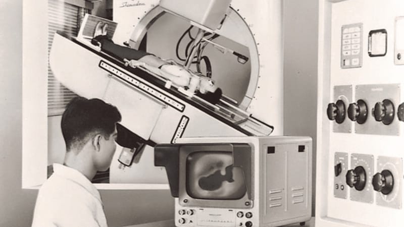
The world’s first Remote-controlled X-ray R/F System was launched in 1961. The achievement of remote control essentially reduced medical practitioners’ radiation exposure to zero. This revolutionary technology would later become the standard, and today it is widely adopted around the world.
At the time, transmitting X-ray fluoroscopic images and displaying them on a monitor was a revolutionary invention. It enabled medical practitioners to remotely operate the system safely from a room separate from the room in which the system was installed, while viewing images efficiently in a bright location. This revolutionary system not only eliminated the risk of radiation exposure, but also drastically reduced the operational burden of examinations.
The spread of the Remote-controlled X-ray R/F System was faster than expected. Each time the system was delivered to hospitals throughout Japan, the number of gastric cancer examinations went up, leading to earlier detection. By 1965, five years after the system was launched, it was reported that the number of gastric cancer cases had started to decline.
On-site Decision Making Continued to Produce Technological Innovation
The history of gastric X-ray examinations using the Remote-controlled X-ray R/F System continued for a long time, and Shimadzu continued to focus on making examinations smoother and more efficient.
Let’s look at a few examples. For instance, when taking still X-ray images, 9 to 11 images are generally required per patient. In the case of a detailed examination, that number can be as high as 20 to 30. When taking the images, each recording film was sandwiched between plates called cassettes, and the films had to be switched after each taking.
Later, a device that could accommodate multiple cassettes was made, but each device could only hold about five. Even though the device could be operated from a separate room, the patient who had to drink the contrast medium would need to wait while the examination was paused to change cassettes. This added time and labor to the examination, mainly from the need to go in and out of the examination room every time the cassettes needed to be changed.
Then in 1971, Shimadzu became the first in the world to commercialize a cassette-less system that could take 100 images continuously by changing films automatically. This was a response to feedback from on-site practitioners who did not want to miss the time frame needed to capture images.
Again in 1990, Shimadzu became the first in the world to launch a radiography table with an elevating function to adjust the height so that patients can easily get on and off the table This function was created in consideration for the elderly and also helped alleviate back strain on technicians, who took hundreds of images each day.
Additionally, in 2004, Shimadzu launched the world’s first direct-conversion flat-panel detector (FPD) that digitized X-ray fluoroscopic images and enabled both video and still images to be taken. This major technological innovation changed history from the age of film to the age of high-quality digital video.
These devices and functions that aimed at ensuring the safety and alleviating the difficulty of treating patients by medical professionals are now the gold standard for medical devices. Their ease of use is broadly acknowledged around the world.
X-ray Diagnosis Continues to Evolve
The appearance of the world’s first Cassette-less R/F System in 1971 launched a “cassette-less era” that streamlined and alleviated the workload of examinations.
Continually listening to feedback from the medical frontline and developing products that foresee what the demand will be, is a tradition at Shimadzu. This tradition has been passed down through the generations. For example, it has led to Shimadzu’s development of devices that enable functions to be flexible, rather than requiring customers to purchase a new device each time a new function is developed. Shimadzu continues to work hand in hand with the medical frontline, not only by contributing to hospital management but also by quickly providing functions that are useful at the frontline.
Sixty years have passed (as of 2021) since the appearance of the Remote-controlled X-ray R/F System, which eliminated radiation exposure to medical practitioners for the first time in the world. Today, X-ray examination technology is used for multiple purposes, including examination for gastric cancer as well as orthopedics, urology, and emergency medicine, and more recently for pancreatic, biliary tract, and liver cancer, and its fields of use continue to expand.
Shimadzu’s product lineup derived from the Remote-controlled X-ray R/F System has grown to more than 20 different models, including the development of devices that support advanced surgery of the brain, as well as blood vessels of the heart, and other circulatory organs. These can be said to come from a history of experiences using customer’s real feedback in product development, and at times conducting joint development with them.
The pursuit of reducing radiation exposure is part of the basic philosophy of Shimadzu’s development of X-ray imaging diagnostic devices. Even now, Shimadzu continues to constantly strive to innovate by actively developing methods for creating clearer images with advanced image-processing technology, and with low doses of radiation.
Moving forward, the use of AI will be key. It will not only make examinations more efficient and further reduce radiation exposure, but will also help improve the level of diagnosis worldwide. This will make diagnosis and treatment using X-rays more convenient, effective and could certainly lead to a future with earlier detection and treatment of disease.

 Copied