
Development of AVS Support System for Primary Aldosteronism
Sampling the adrenal vein is an effective means of determining a treatment plan for primary aldosteronism, but
it can take several days to obtain results. Furthermore, samples are acquired from multiple locations. That has resulted in
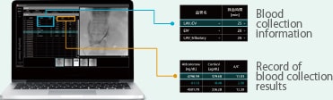
problems due to the difficulty in integrating image information with measurement results and managing the data.The AVS support
system for primary aldosteronism marks adrenal vein images with the locations where blood was acquired from adrenal veins during
sampling and displays measurement results linked to those marked locations. Consequently, the system enables an accurate
understanding of measurement results at each adrenal vein location from only a glance.
- Primary Aldosteronism
- Primary aldosteronism causes high blood pressure due to excessive excretion of the hormone aldosterone from the adrenal gland. About 5 to 10 % of high blood pressure cases are said to be caused by primary aldosteronism, which can be resolved in nearly 50 % of cases through adrenal gland surgery.
AVS Support System for Primary Aldosteronism
-
(1)Insert a catheter into the adrenal vein for venography.
-

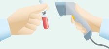
(2)Based on an X-ray image, insert a catheter in the adrenal vein to acquire blood at
multiple locations and read the sample ID information from each blood collection tube. -
(3)Use the sample ID information to record the blood collection
location information on the X-ray image.-
・Angiography System that Reads Sample ID Information from Blood Collection Tubes and Links it to Blood Collection Locations on X-Ray Images
Registers sample ID information from blood collection tubes and the corresponding blood collection locations.
-
-
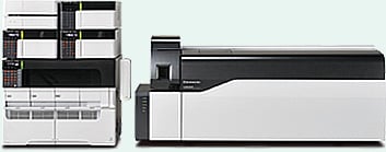
(4)Read the sample ID information from each blood collection tube and
analyze the samples by mass spectrometry.-
・Mass Spectrometer for Quantitating Aldosterone and Cortisol Levels in the Blood (Research Use Only)
Quickly and accurately measures a wide variety of substances from a small quantity of blood samples.
-
-
(5)Use the sample ID information to associate the LC-MS/MS analysis results with each
blood collection location and then prepare an AVS examination report.-
・Software for Displaying Measurement Results Associated with Blood Collection Locations on X-Ray Images
X-ray images (morphology information) with different characteristics can be integrated with measurement results (biological information) and managed as a single set of information.
-
Note: This system is intended for research use (scheduled for release only in Japan).
FOR RESEARCH USE ONLY. NOT FOR USE IN DIAGNOSTIC PROCEDURES.
Collaborative Research Partner
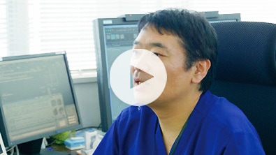
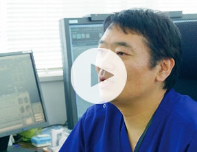
- Tohoku University Hospital
- Dr. Kei Takase
“Using a combination of diagnostic imaging technology and measurement technology for component analysis, the AVS support system enables faster and more accurate diagnostic capabilities. The ability to determine, on-the-spot from a localization diagnosis, where the problem is located in the adrenal vein will lead to less invasive treatments in the future. This type of sampling technique can be used not only for the adrenal gland but also for various other endocrine organs and endocrine disorders. Therefore, I think that our development experience can also be deployed for other endocrine disorders in the future.”