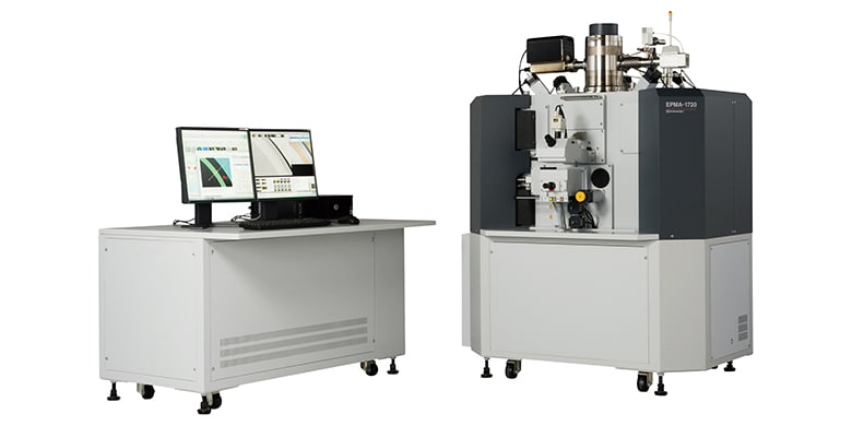
EPMA-1720
- Enables highly sensitive measurement of the distributions of fluorine, magnesium, and other trace elements on the tooth surface. - Possible to analyze the irregularly shaped, extremely uneven tooth surface without sample preparation. - Can be used in research on the tooth growth process and prevention of tooth decay.
Human teeth can be divided into the crown, which is visible in the mouth, and the root, which is hidden by the gingiva. The crown is covered with enamel and is the hardest tissue in the human body. Approximately 95 % of enamel is an inorganic substance consisting mainly of hydroxyapatite (Ca10(PO4)6(OH)2). Cavities (dental caries) occur when the amount of decalcification of the enamel caused by acids produced by caries-causing bacteria exceeds the amount of recalcification. It is known that coating teeth with fluoride or use of the artificial sweetener xylitol is effective for promoting recalcification. Analysis of trace elements is extremely important in research on this tooth growth process and prevention of tooth decay. This article introduces an example of an analysis of the surface of a deciduous molar that was regularly coated with fluoride by a dentist, in which a mapping analysis was conducted using a Shimadzu EPMA-1720HT EPMATM electron probe microanalyzer with stage height correction (trace mapping analysis function of the EPMA) following the uneven surface topography of the tooth.
March 1, 2022 GMT