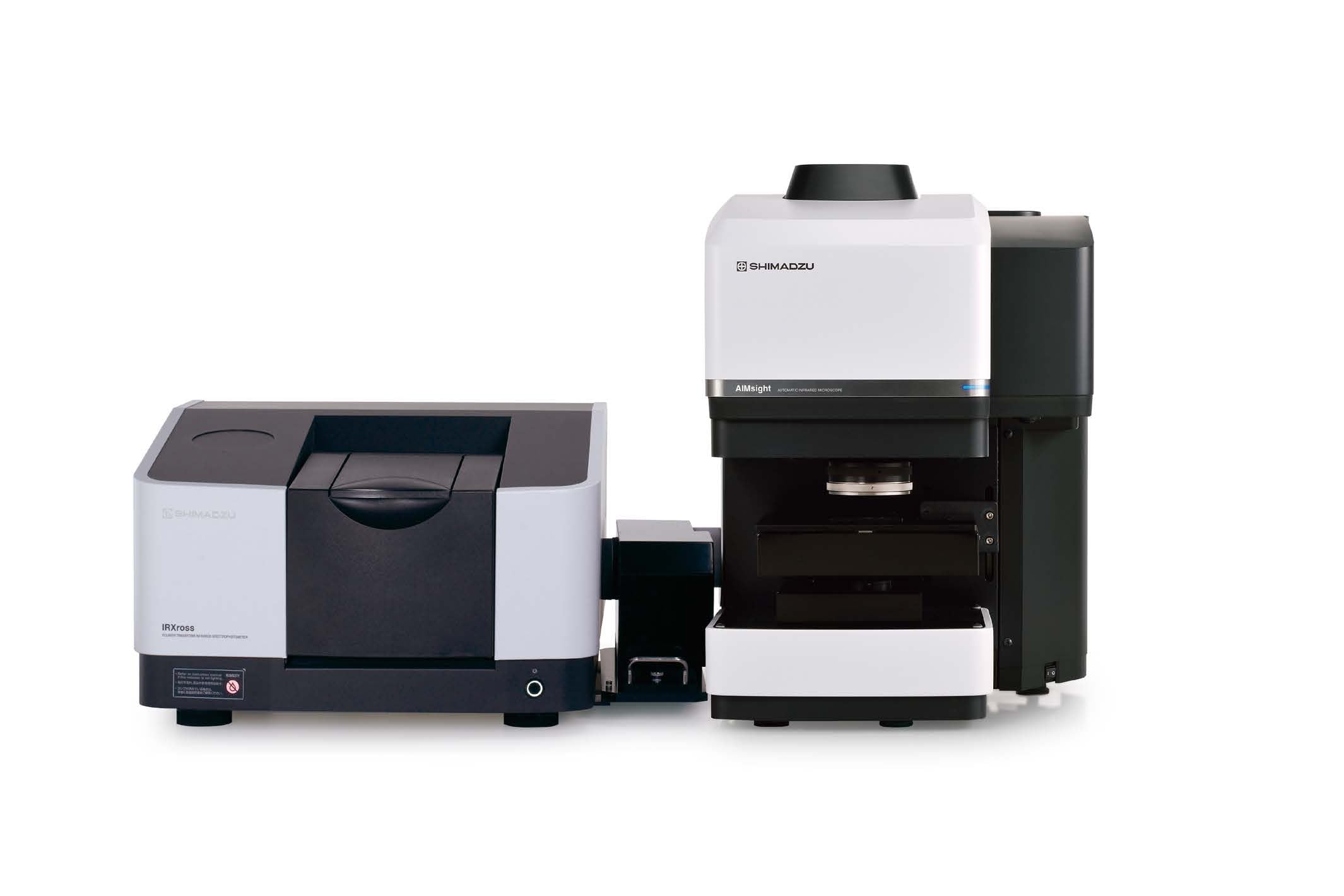
AIMsight™



INDUSTRY
Microplastics
KEYWORD
Microplastics, particle analysis
INTRODUCTION SERVICE&PRODUCTS
AIMsight™
The prevailing belief is that the majority of microplastics (MPs) and other marine plastic waste is generated inland, where it undergoes physical and chemical degradation as it travels along waterways until it finally flows into the ocean. However, the details of this dynamic process are not well understood. In particular, infrared microscopes are used to analyze smaller MPs less than 1 mm in size. We interviewed Associate Professor Tomoya Kataoka of the Department of Civil & Environmental Engineering at Ehime University, whose work developing new models that use geometric relationships to correlate the projected area and mass of MPs has attracted interest from MP researchers around the world.

Department of Civil & Environmental Engineering, Ehime University
Tomoya Kataoka, Associate Professor
*The affiliation and title of the customer are correct as of the time of publication.
*Affiliates and titles of the interviewee are current as of the time of reporting.
Kataoka Laboratory
URL
https://www.cee.ehime-u.ac.jp/~tkata/index.html
My main research topic is the dynamics of MPs in inland waters. Throughout the world, MP monitoring studies have mainly collected MPs using nets with a 300 μm mesh. Assuming that the number of plastic particles increases as they break down into smaller pieces, there should be greater numbers of smaller MPs. However, the size distributions of MPs observed in rivers to date contradict this assumption, showing fewer MPs below 1 mm than would be expected (Fig. 1). This inconsistency arises because MPs smaller than the mesh size of the collection net pass through it, so the numbers collected do not accurately represent the numbers present in the water (Fig. 1). We focus on field surveys targeting these smaller MPs below 1 mm which have previously been overlooked.
A major challenge in understanding the dynamics of smaller MPs below 1 mm is mass measurement. MPs larger than 1 mm that are caught by a typical sampling net can be weighed individually on an ultra-microbalance, but smaller MPs cannot be reliably weighed directly in this way. However, after analyzing large amounts of empirical data, we found a strong correlation between the projected area (the area when viewed from directly above) of an MP and its mass. We were also able to explain this correlation theoretically using geometric relationships based on the morphological characteristics of MPs. Other researchers have previously proposed mass conversion formulas that calculate MP mass from a volume estimated using an assumed thickness, but these formulas tend to overestimate the mass of MPs. In contrast, the geometric correlation models we established are based on actual measurements of MPs collected in the field. This new mass estimation method has enabled more accurate estimation of the mass of smaller MPs.
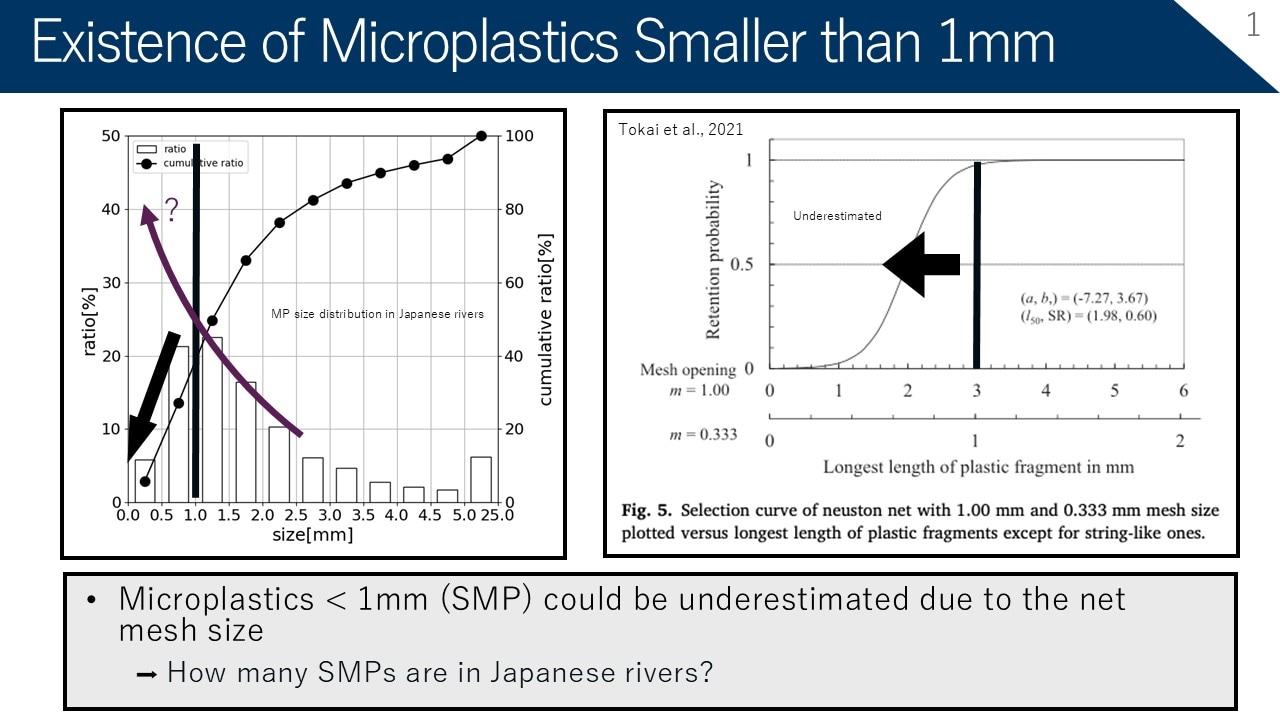
Fig. 1 Size Distribution of MPs Collected from Rivers
Quantitative analysis of MPs by FTIR and IR microscopy has typically been based on counting the number of MPs. However, when assessing environmental dynamics and physiological effects, we also need mass and volume data and an efficient means of obtaining them. We hoped the correlation model we developed could be integrated into the particle analysis software for infrared microscopes, as this would allow the mass and volume of smaller MPs to be calculated automatically. As we have used Shimadzu’s FTIR and IR microscope instruments for many years and have been exchanging information with Shimadzu’s Global Environmental Management Department on technical issues in MP monitoring for the past several years, I had the opportunity to propose this idea for the particle analysis software to Shimadzu.
We are delighted that our models have been incorporated into the particle analysis software for Shimadzu’s IR microscopes, which are used around the world. The software automates the particle characterization process, including MP identification by FTIR or IR microscopy, measurement of projected area, and estimation of mass and volume. This greatly reduces the labor involved in MP analysis as well as human error, and can substantially improve overall research efficiency. Furthermore, because the software will come standard with the same correlation models both in Japan and internationally, anyone, wherever they are located, will be able to produce objective estimates of MP mass.
This type of method harmonization makes it easier to compare data between research groups and could play an important role in accelerating the elucidation of MP dynamics on a global scale.
We typically collect smaller MPs by sampling the water itself (around 3 to 5 liters). Sampling nets are primarily used to collect MPs of 1 mm or larger, but for smaller MPs, nets with finer mesh become clogged, which causes problems.
Instead, we either collect river water with an aluminum bucket from a bridge and bring the water back to the laboratory in an aluminum pouch, or use a pump to pass water through a filter and bring the filter back for analysis. The bucket is used to investigate the concentration of smaller MPs at the river surface, and the pump is used to investigate the concentration of MPs deeper in the river. The amount of water collected by the bucket or pump is just one five-thousandth of the volume that passes through a typical sampling net (on average, 15 m3), but given that there are 3 to 4 orders of magnitude more MPs under 1 mm than over 1 mm in size, this volume is sufficient for analysis. In fact, we have to be careful not to collect too much water, as this can complicate subsequent processing.
Samples brought back to the laboratory are processed and analyzed as shown in Fig. 2. First, the samples are filtered through a nitrocellulose filter (pore size: 5 µm).
Next, the filter is dissolved, contaminants are decomposed by oxidation, and low-density particles are isolated centrifugation.
Finally, the resulting particles are separated using filters with two different pore sizes (100 µm and 26 µm): the particles retained by the 100 μm filter are analyzed by FTIR, and the particles retained by the 26 μm filter are analyzed by IR microscopy.
Preventing contamination is an important part of this process. Samples are processed in a clean booth, and analysis is carried out very carefully, including blank checks. Using this method, we have been identifying the materials of MPs retained on the 26 μm filter by point measurement with IR microscopy. However, the smaller the particles, the more of them are retained on the filter, and the longer analysis and characterization take. I have heard that Shimadzu’s particle analysis software automatically determines projected area and volume, which helps reduce the time spent on mapping measurements. We look forward to using the area measurement capabilities of the new particle analysis system to analyze samples more efficiently.
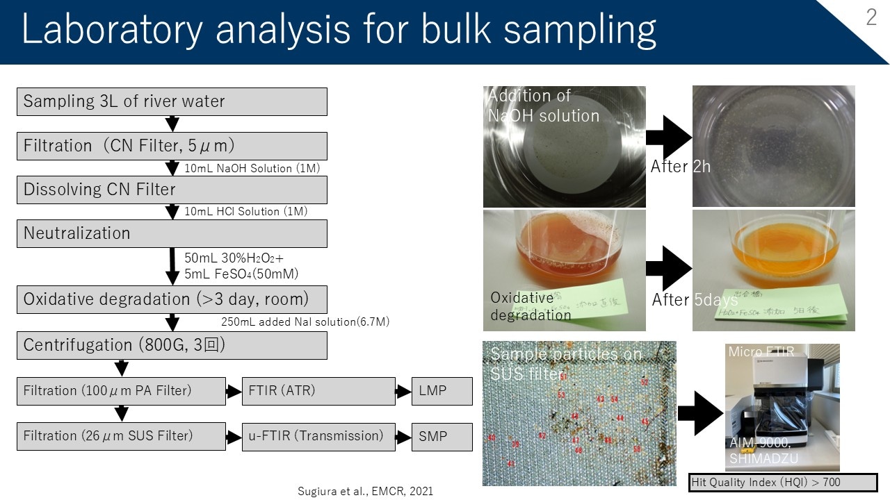
Fig. 2 Sample Treatment Process
I would like to see further improvements in the functionality and accuracy of the particle analysis software. Specifically, integrating AI into particle image processing should enable faster and more efficient analysis, so we hope Shimadzu will pursue development in this area. Because AI requires a graphics processing unit (GPU) in the analysis computer, integrating AI would also require a review of hardware specifications, but it should lead to faster and more accurate material identification and analysis.
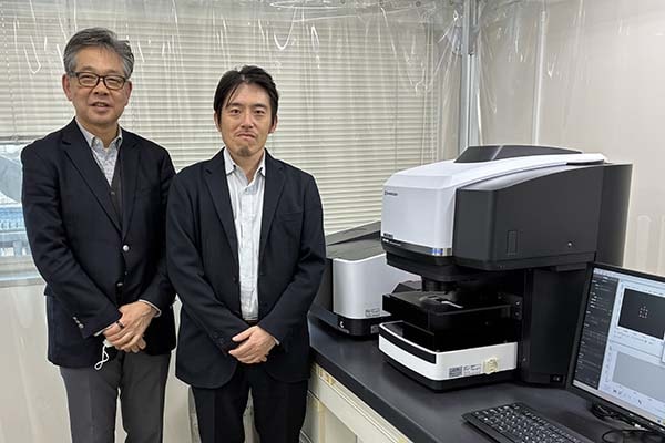
Associate Professor Tomoya Kataoka uses an infrared microscope and a number of other Shimadzu products in his laboratory. We are grateful for the valuable insights provided in this interview.
*The product names in this article are correct at the time of publication.