Observation of Lamellar Structure of Polymer Film (SPM)
Polymers are utilized all around us in a variety of forms. The surfaces of these polymer items have an intriguing form typified by a lamellar structure, which affects the transparency and strength of the material.
In this example, the surface of a polyethylene film sample was observed using a scanning probe microscope, and a 3D image and elasticity image were simultaneously obtained.
The tiny bumps on the surface are visible in the 3D image. The elasticity image clearly shows the lamellar structure and the crystallization of the polymer. (The more the crystallization advances to increase the hardness, the lighter the area appears.)
The images represent an area 1 µm long on each side. In this way, an SPM can be used to easily obtain important information about polymer films under atmospheric conditions.
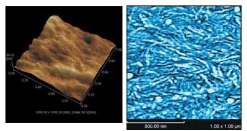
3D Image (Left) and Elasticity Image (Right) of Surface of Polyethylene Film
*For examples of observation data obtained using an SPM, click here.
Scanning Probe Microscope
A scanning probe microscope (SPM) scans the sample surface with a microscopic probe to provide high-magnification 3-dimensional observations. It permits nm-order observations and shape measurements of solid and film surfaces in air or in solution.