| Description | Q'ty |
|---|---|
| USF-2000A W/O AIRCOMP | 1 |
| COMPRESSOR,ELP86-15T | 1 |
| DISPLACEMENT DEVICE CE (2MM) | 1 |
| Extensometer Calibration Devices | 1 |
| NR-500 AD BNC SET | 1 |
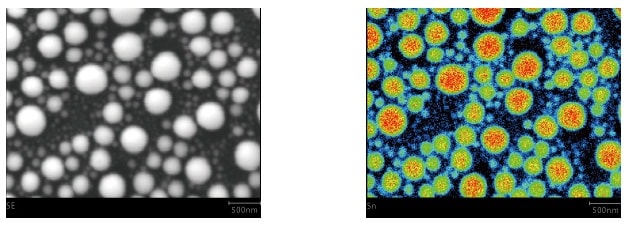
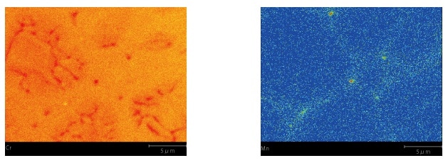
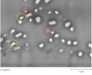
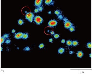
| EPMA-8050G 4CH | 1 |
| EPMA-8050G PC ASSY | 1 |
| EPMA-8050G IP BACKUP POWER SUPPLY ASSY | 1 |
| AIR COMPRESSOR,PC3-5.5TGL | 2 |
| THERMO-CON,HEC002-A5B S | 1 |
| Description | Q'ty |
|---|---|
| ELECTRON PROBE MICROANALYZER EPMA-8050G 4CH | 1 |
| EPMA-8050G PC ASSY E | 1 |
| EPMA-8050G IP BACKUP POWER SUPPLY ASSY | 1 |
| THERMO-CON,HEC002-A5B S | 1 |
| COMPRESSOR,PC3-5.5TGL | 2 |
| Description | Q'ty |
|---|---|
| ELECTRON PROBE MICROANALYZER EPMA-8050G 4CH | 1 |
| EPMA-8050G PC ASSY | 1 |
| EPMA-8050G IP BACKUP POWER SUPPLY ASSY | 1 |
| AIR COMPRESSOR,PC3-5.5TGL | 1 |
| THERMO-CON,HEC002-A5B S | 1 |
| AIR COMPRESSOR,PC3-5.5TGL | 2 |
| Description | Q'ty |
|---|---|
| ELECTRON PROBE MICROANALYZER EPMA-8050G 4CH | 1 |
| EPMA-8050G PC ASSY | 1 |
| EPMA-8050G IP BACKUP POWER SUPPLY ASSY | 1 |
| AIR COMPRESSOR,PC3-5.5TGL | 2 |
| THERMO-CON,HEC002-A5B S | 1 |
| Description | Q'ty |
|---|---|
| ELECTRON PROBE MICROANALYZER EPMA-8050G 4CH | 1 |
| EPMA-8050G PC ASSY | 1 |
| EPMA-8050G IP BACKUP POWER SUPPLY ASSY | 1 |
| AIR COMPRESSOR,PC3-5.5TGL | 2 |
| THERMO-CON,HEC002-A5B S | 1 |
| Description | Q'ty |
|---|---|
| ELECTRON PROBE MICROANALYZER EPMA-8050G 4CH | 1 |
| EPMA-8050G PC ASSY | 1 |
| EPMA-8050G IP BACKUP POWER SUPPLY ASSY | 1 |
| AIR COMPRESSOR,PC3-5.5TGL | 2 |
| THERMO-CON,HEC002-A5B S | 1 |
| Description | Q'ty |
|---|---|
| ELECTRON PROBE MICROANALYZER EPMA-8050G 4CH | 1 |
| EPMA-8050G PC ASSY | 1 |
| EPMA-8050G IP BACKUP POWER SUPPLY ASSY | 1 |
| AIR COMPRESSOR,PC3-5.5TGL | 2 |
| THERMO-CON,HEC002-A5B S | 1 |
| Description | Q'ty |
|---|---|
| ELECTRON PROBE MICROANALYZER EPMA-8050G 4CH | 1 |
| EPMA-8050G PC ASSY E | 1 |
| EPMA-8050G IP BACKUP POWER SUPPLY ASSY | 1 |
| THERMO-CON,HEC002-A5B S | 1 |
| COMPRESSOR,PC3-5.5TGL | 2 |