Q: I get extremely high noise with microscope transmission measurements. What is it caused by?
A : Poor adjustment of the condenser mirror is one cause of high levels of noise during microscope transmission measurements. During microscope transmission measurements, the light may not be focused even if foreign matter is clearly apparent in the visible image. In this case, adjust the condenser mirror under the sample stage. Normally, during microscope installation, the condenser mirror is adjusted to focus the light onto the MCT detector element when no aperture plate is placed on the sample stage. (See Fig. 1 left-hand diagram.) When measurements are performed with the aperture plate in position, the light path length changes due to the different refractive index of the aperture plate material, such that the focal point becomes displaced as shown in the right-hand diagram in Fig. 1. (The refractive index of air is 1.0, but 2.38 for the diamond cell or 1.42 for a BaF2 window.) If measurements are performed in this situation, the light intensity drops and the noise increases in the resulting spectrum.
In Shimadzu infrared microscopes, when a pinhole is observed in the visible image (Fig. 2 left-hand image), the light is correctly focused and the optical axis aligned. This achieves optimal sensitivity.
Fig. 3 shows a comparison of spectra when the light is correctly and incorrectly focused. The sample is trace foreign matter on a diamond cell. It was measured using a 10 × 10 µm aperture size. A comparison of the two spectra indicates extremely high levels of noise when the light focus is not correctly adjusted.
The adjustment method described above differs from instrument to instrument. Consult the manufacturer of your infrared microscope, if you use a non-Shimadzu product.
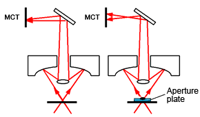
Fig. 1 Light Path for Microscope Transmission Measurements
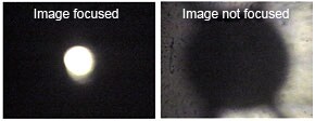
Fig. 2 Adjusting the Pinhole on the Visible Image
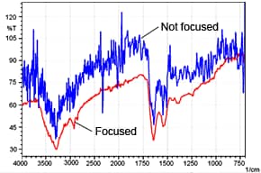
Fig. 3 Microscope Transmission Spectra of Foreign Matter