SPM-Nanoa - Applications
Scanning Probe Microscope/Atomic Force Microscope
Applications
| Applications | Date Creation Date |
|---|---|
2025-07-22 | |
2025-07-22 | |
2024-06-04 | |
2023-10-24 | |
2023-04-12 | |
2022-05-16 | |
2022-04-05 | |
2022-01-06 | |
2021-09-24 | |
2021-09-24 | |
2021-09-24 | |
2021-09-24 | |
2021-07-16 | |
2021-05-06 | |
2021-04-01 |

Most of the documents on the LITERATURE is available in PDF format. You will need Adobe Acrobat Reader to open and read PDF documents. If you do not already have Acrobat Reader, you can download it free at the Adobe's Website. Click the GET ADOBE READER icon on the left to download a free copy of Adobe Acrobat Reader.
Application Software
Application software for a wide variety of samples, from soft to hard materials, can provide powerful help for observing what you want to observe.
| Hard Materials | ・Nanoparticles ・Nanofibers ・Fillers ・Ceramics ・Metals |
|---|---|
| Soft Materials | ・Plastics ・Rubbers ・Films ・Biological materials ・Composite materials |
| Life Sciences and Healthcare | ・Lipid membranes ・Cells ・Biological molecules ・Hair |
| Electronics | ・Battery materials ・Semiconductors ・Recording media |
What do you want to observe?
* Option
Hard Materials
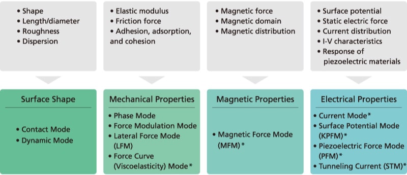
Silica Nanoparticles
Observation of silica nanoparticles confirmed uniformity of nanoparticle sizes.
Life Sciences and Healthcare
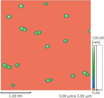
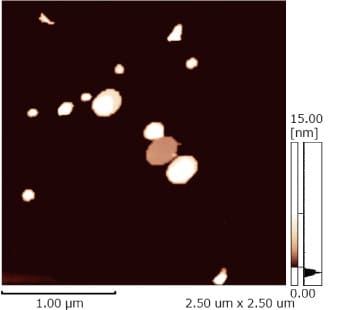
Extracellular Vesicles
The large particles shown in the center are extracellular vesicles. With the ability to not only observe shapes, but also evaluate mechanical properties, the system is expected to be useful for identification and Drug Delivery System (DDS) research for exosomes, liposomes, and typical polymer micellization pathogens, and other applications (using Nano 3D Mapping Fast).
Soft Materials
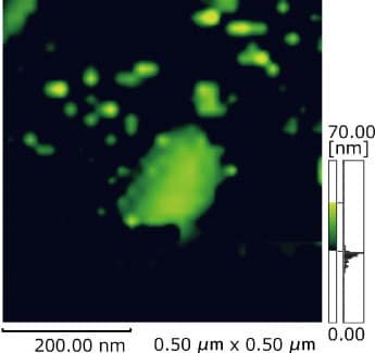
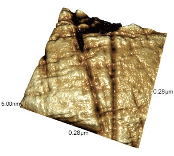
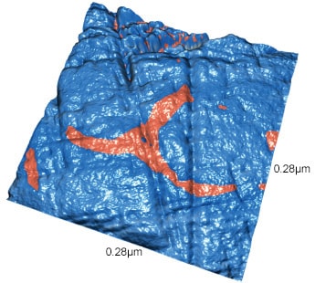
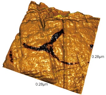
PVP/CNF Composite Materials
Surface Shape
Phase
Surface Shape + Phase
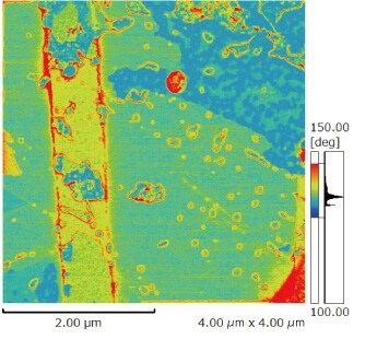
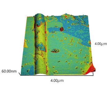
A water mixture of cellulose nanofiber (CNF) and polyvinylpyrrolidone (PVP) was observed electrospun onto a silicon substrate. The surface shape image shows the cylindrical shape of the fibers and the phase image shows physical property differences of CNF and PVP fibers as differences in contrast.
(Sample source: Professor Nakai, Graduate School & Faculty of Bioresources, Mie University)
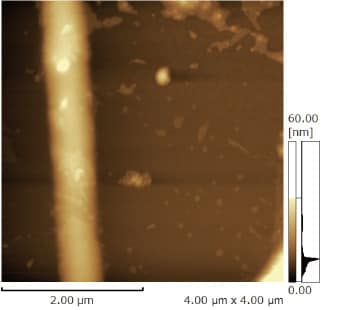
Lipid Membranes
Surface Shape
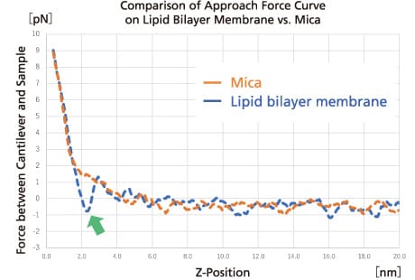
A patch-shaped lipid membrane about 6 nm thick was observed (arrow) near the center of the surface shape image (left). The force curve acquired from on top of the lipid membrane (right) indicates the variations in force generated as the probe penetrated the membrane.
Electronics
Single BaTiO3 Crystal
Surface Shape
Phase
Surface Shape + Phase
BaTiO3, a strong dielectric, was observed using the piezoelectric force mode (PFM).
The amplitude and phase images clearly show the polarized domain structure.

