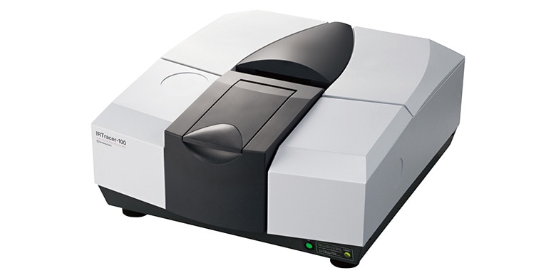
IRTracer-100
Spectrophotometric Analysis
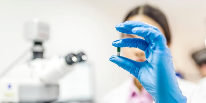
Amyloid-β is a peptide that consists of approximately 40 amino acid residues. Amyloid fibrils (fibrous aggregates), which occur as a result of the formation of parallel β sheets by intermolecular association of amyloid-β, are the main component of the senile plaques (amyloid plaques) seen in the brains of Alzheimer’s disease patients. Moreover, the formation mechanism and structure of amyloid fibrils have attracted great interest, as amyloid fibrils are also implicated in other neurodegenerative diseases, including Parkinson’s disease. With FTIR, it is possible to evaluate the aggregation of amyloidβ by analyzing the amide I band around 1,650 cm-1, which originates from stretching vibrations of the C=O group of the peptide bond. Fig. 1 shows the peptide bond structure. The secondary structures (α-helix, β-sheet, β-turn, random coil, and other local 3-dimensional conformational features) of the amyloid-β peptide can be obtained by curve fitting (peak splitting). Curve fitting is method in which the waveforms of each absorption band are expressed by an approximate curve such as a Lorentzian curve or Gaussian curve, and the peak information (position, intensity, full width at half maximum) of the approximate curve of each absorption band is optimized so as to minimize the difference between the calculated spectrum and the measured spectrum (2). This article introduces the results of an evaluation of amyloid-β aggregation.
June 8, 2020 GMT