MALDI-8020
- Simple imaging analysis of peptides and proteins in the rat brain on an affordable, easy-to-use benchtop MALDI-TOF system - High quality spectra with class-leading mass resolution and sensitivity producing detailed MALDI images - Workflow applicable to many tissue types and with different methods of matrix application
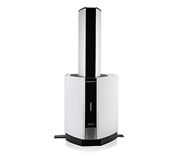
MALDI mass spectrometry imaging (MSI) is a powerful technique that utilizes the capabilities of the spectrometer to collect thousands of individual spectra at various positions on a sample. Subsequently, using dedicated software, the resulting detected ions can be spatially represented as pseudo-colour images to visualize key molecules relative to their position in the sample / tissue. The MALDI-8020 linear time-of-flight (TOF) mass spectrometer (Fig. 1) is capable of generating quality MS images, such as lipids in rat brains. The ability to rapidly exchange sample plates in the instrument (<3 mins) and the short ‘instrument-ready’ time mean that acquisitions can be quickly started, which is advantageous during optimisation of imaging methods. We have previously demonstrated MALDI imaging for a variety of target analytes on the MALDI-TOF benchtop system with the analysis of fingerprints, soybeans and PET films (Shimadzu Application News; 01-00389-EN and 01-00392-EN). Here, we demonstrate the capability of our imaging platform to easily achieve quality mass spectra and MS images for intact proteins from tissue sections and for peptides following on-tissue digestion, with linear mode analysis of rat brain tissue.
January 26, 2023 GMT
Some products may be updated to newer models