Cell Pocket™
- Cell Pocket provides a simple means of quantifying sensuous observations. - Quantified observation information can be used for objective evaluation of culture conditions. - Objective assessment results can be shared with lab members along with data.
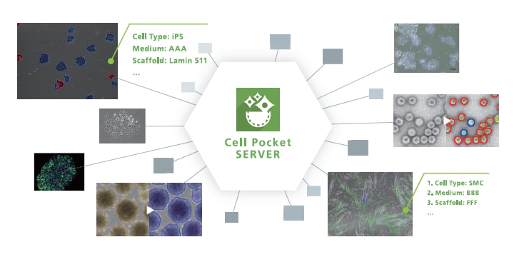
Human pluripotent stem cells are a heterogeneous population that change constantly during the culture process. The changing morphology of human pluripotent stem cells is considered an important quality characteristic, and evaluating product quality at the completion of manufacture, as is the case with small molecule drugs, is not a suitable approach for cell preparations. Given this backdrop, the industrialization of regenerative medicine and cell therapy requires methods of evaluating the status of cells that are noninvasive and provide observations over time. A well-known noninvasive technique is the assessment of cell morphology by image analysis. Analyzing cell morphology once posed a significant challenge, but thanks to dramatic improvements in machine learning techniques and the increased availability of these techniques, cell morphological analysis is now feasible and attracting interest. Shimadzu used segmentation by deep learning, a machine learning technique, to develop Cell Pocket, a web application that offers a simple platform for image analysis. Cell Pocket is a user-friendly advanced image analysis tool that allows users to quantify their sensuous or experimental observations and derive important findings based on these observations. This article presents an example analysis that visualizes changes in spheroid morphology during the differentiation and maturation process of spheroids of endoderm cells derived from human iPSCs.
March 28, 2023 GMT