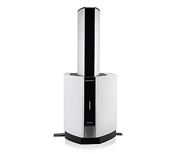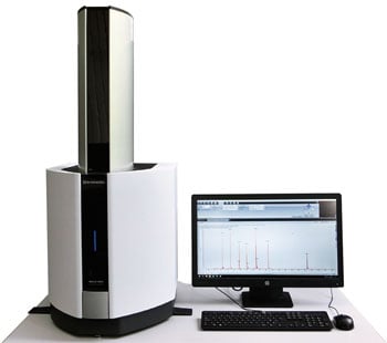
MALDI-8020
- Simplified protein imaging analysis on an affordable benchtop MALDI-TOF system. - Integration of immunohistochemistry into the MALDI imaging workflow (MALDI-IHC) for targeted analysis. - Multiplex analysis allowing visualization of 20 proteins in a single imaging run.
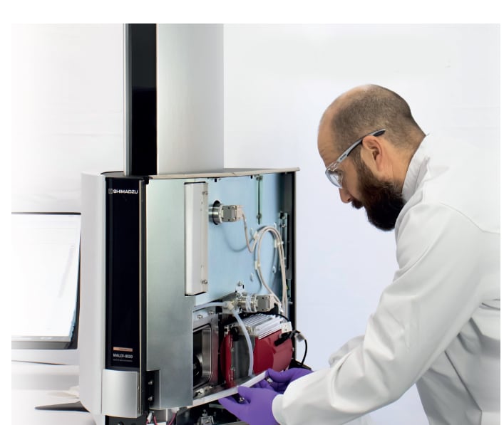
Here, we demonstrate an entry level MALDI imaging solution to allow the precise mapping of proteins usingMiralysMALDI-IHCimagingprobes(AmberGen, MA, USA). Matrix assisted laser desorption ionization immunohistochemistry (MALDI-IHC) is a technique which uses a photocleavable peptide mass tag conjugated with an antibody to label proteins within a tissue sample. Subsequent cleavage of the mass tag allows for this to be detected by MALDI-TOF. Using mass spectrometry imaging (MSI), spatial information of the cleaved peptide can be determined and, from this, the locations of the antibodies, and hence the target proteins in the tissue, can be inferred. Through MALDI-IHC, we can further our knowledge of cellular interactions which is critical to the development new treatments and the Shimadzu benchtop MALDI instruments are ideal platforms to begin exploring the world of MALDI-IHC. In this study, we have analysed FFPE human tonsil sections using both a 6-plex and 20-plex antibody probe panels at a lateral resolution of 30 μm, demonstrating a reliable low-cost approach with good quality results.
February 2, 2025 GMT
Some products may be updated to newer models