

2. Differences from Mass Spectrometry-Based Amino Acid Sequence Analysis
- 1. What Is a Protein Sequencer?
- 2. Differences from Mass Spectrometry-Based Amino Acid Sequence Analysis
- 3. Example of a Complete Peptide Sequence Analysis Using Complementary Application of ISD and the PPSQ Protein Sequencer
Amino acid sequence analysis can be performed either using a protein sequencer based on Edman degradation or with a mass spectrometer in combination with a search engine. The analytical workflows for each approach are outlined below.
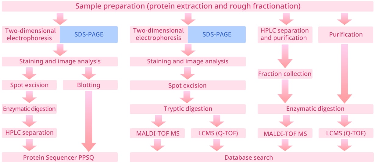
Features of Edman Degradation-Based Amino Acid Sequencing
- Well suited for sequential identification from the N-terminal amino acid of a protein and for confirming sequence homogeneity.
- Intact proteins can be submitted directly for analysis, enabling long-chain sequence determination.
- Amino acids with identical mass (leucine and isoleucine) or similar masses (lysine and glutamine) can be readily distinguished and identified.
- Identification is possible even for unknown proteins not registered in existing databases.
Features of Mass Spectrometry-Based Amino Acid Sequence Analysis
- High throughput.
- Results can be obtained from trace amounts of sample.
- Edman degradation addresses the limitations of mass spectrometry-based amino acid sequence analysis.
- Combining mass spectrometry with Edman degradation yields more accurate and information-rich sequence data.
2.1 Peptide Mass Fingerprint (PMF) Method
-
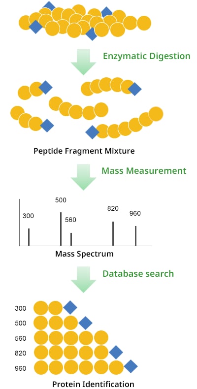
With the peptide mass fingerprint (PMF) method, a purified single protein is subjected to enzymatic digestion, generating a mixture of multiple peptides (enzymatic digests) from a single polypeptide chain. The mass of each digest in the mixture is measured using a mass spectrometer, such as a matrix-assisted laser desorption/ionization time-of-flight mass spectrometer (MALDI-TOF MS). The resulting masses are searched against a database to assign them to proteins in the database, from which the primary structure of the protein is inferred. -
2.2 In-Source Decay (ISD) Method
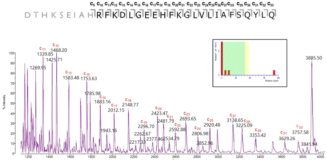
This method uses a matrix-assisted laser desorption/ionization time-of-flight mass spectrometer (MALDI-TOF MS) to analyze an isolated and purified protein using a matrix that promotes fragmentation. The resulting ions generated by ISD can be detected, enabling sequence analysis of intact proteins without the need for tryptic digestion.
An example of BSA analysis by the ISD method is shown here. With this approach, simply increasing the laser irradiation power after molecular weight measurement enables straightforward internal sequence analysis of high-purity proteins and peptides.
2.3 MS/MS Ion Search
-
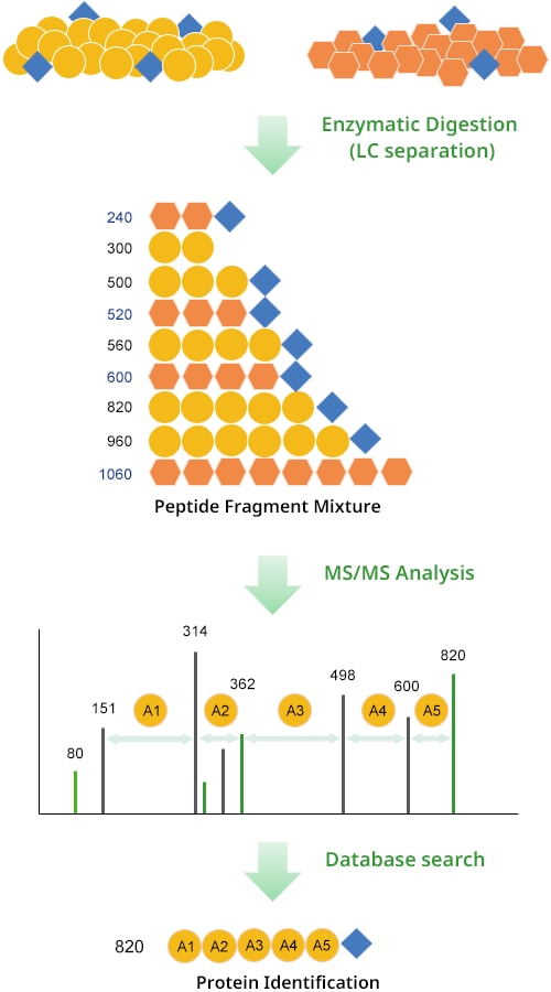
Unlike the PMF method, this approach generates a mixture of multiple enzymatic digest peptides by subjecting a protein mixture as opposed to a single protein to enzymatic digestion. The resulting digest mixture is first separated by high-performance liquid chromatography (HPLC). Each separated peak is then measured by a mass spectrometer, such as a quadrupole time-of-flight LC-MS/MS (Q-TOF). During this analysis, in addition to the mass of each enzymatic digest, the masses of fragment ions generated by collisions with gas molecules (equivalent to MS/MS data) are also measured. The resulting data are used to conduct a database search, and the identified peptides are matched to proteins in the database to identify the protein(s) of interest.
-