iMLayer
- Label free image analysis of organoids samples. - Easy setting up of experiments using Shimadzu’s Total Solutions for MALDI Imaging. - Simple data processing possible with IMAGEREVEAL MS.
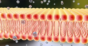
Matrix-Assisted Laser Desorption/Ionization (MALDI) Imaging Mass Spectrometry (IMS) is transforming organoid research by providing spatially resolved molecular insights. Organoids—three-dimensional cellular models derived from stem cells that replicate the structure and function of human tissues—are powerful tools for disease modeling, drug screening, and personalized medicine. MALDI Imaging enhances these applications by enabling the spatial analysis of biomolecules such as proteins, lipids, and metabolites, offering a detailed functional understanding of biochemical processes within complex tissue-like structures. This spatial mapping is particularly valuable for studying conditions like cancer, where tissue heterogeneity significantly influences disease progression and drug response. In this study, we established a standardized workflow using Shimadzu’s complete imaging solution to analyze human lung organoids. Sample preparation was carried out using the iMLayer and iMLayer AERO systems to achieve reproducible and homogeneous matrix deposition. Imaging was conducted with the iMScope QT, and data were processed using IMAGEREVEAL MS. The integration of these tools enabled high-resolution MALDI Imaging with enhanced sensitivity, providing clear lipid distributions within differentiated organoid tissues. Looking ahead, the clinical potential of MALDI Imaging is immense—especially in the context of personalized medicine. As patient-derived organoids become increasingly prevalent, the ability to map molecular distributions at high spatial resolution will provide important insights into patient-specific molecular profiles.
September 29, 2025 GMT
Some products may be updated to newer models