Xctal 5000 - Features
Phase-Contrast X-Ray CT System
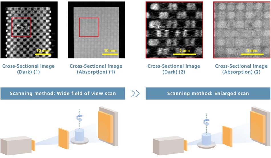
Both Wide Field of View and Detailed Structural Observations
Detailed Structural Observations
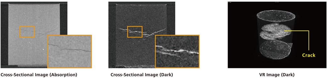
A dark-field image is suited to observations of fine structure, including cracks and the flow of fiber bundles, over a wide field of view.
In contrast, an absorption image is suited to enlarged observations of shapes in detail within samples.
The dark-field image obtained by scanning over a wide field of view makes it possible to ascertain fine structure in the sample as a whole, and to estimate its position.
The absorption image obtained by enlarged scanning makes it possible to observe the shape of fine structure in detail.
When observing fine structure, there is no need to scan repeatedly over a narrow field of view.
Example: Cross-Sectional Images of a CFRP Cloth Material with Cracks
Observations of Fiber Orientation
-
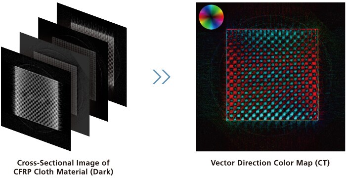
- The flow of fiber bundles, derived from multiple dark-field images, can be aligned with the orientation angle and displayed in color (vector direction color maps). The flow of fiber bundles can be understood intuitively by representing it in color.
Applications (Dark-field Images)
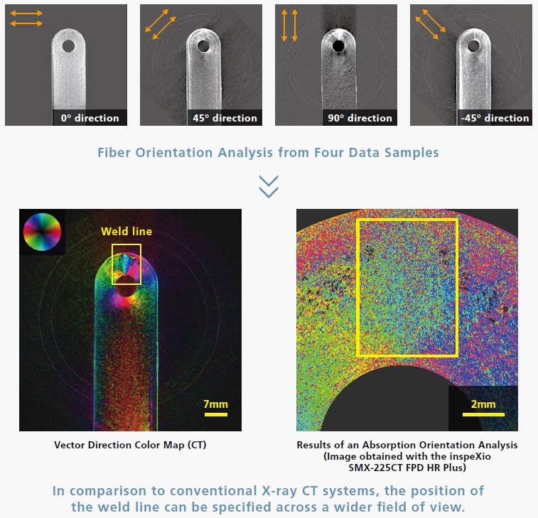
GFRP Injection Molded Items
In injection molding, molding defects can occur when a linear mark (weld line) forms where the molten resins meet.
With this system, dark-field images can be obtained while changing the angle of the sample, so the fiber flow can be analyzed, enabling the position of weld lines to be specified.
Biocoke
Biocoke is a solid biofuel formed from plants through photosynthesis. In this example, fine cracks produced during formation can be visualized using dark-field images. Fine cracks invisible with conventional X-ray CT can be visualized by detecting the scattering of X-rays at the borderline between the material and the crack (air).
The quantitative analysis of microscopic cracks using this system has become a standard evaluation to check whether the combustion properties of biocoke are as close as possible to those of coal coke, thus laying the foundations for achieving a carbon neutral society through conversion to renewable energy.
(Samples and comments provided by: Professor Tamio Ida of the Bio-Coke Research Institute, Kindai University)
The affiliation of the providers of the samples and comments is as of July 27, 2022.
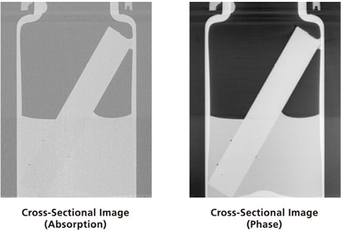
Dual Observation Method for Absorption and Phase
High-Contrast Observations by Absorption Images and Phase Images
A phase image is suited to observations of samples with large density differences.
In contrast, an absorption image is suited to observations of samples with large differences in absorption coefficient.
The X-ray absorption coefficient makes it possible to visualize samples with no contrast differences, as long as there are density differences. Using these two observation methods widens the scope of observations by enabling high-contrast sample observations.
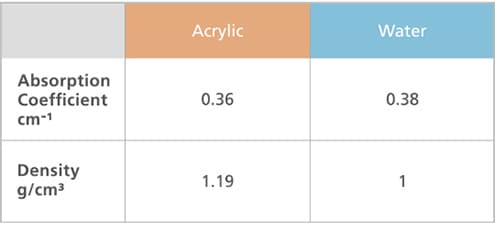
Water and Acrylic
The density difference is larger than the absorption coefficient difference, enabling high-contrast phase image observations.
High-contrast phase image observations are possible if the density difference is approximately 0.2 g/cm3 or larger.
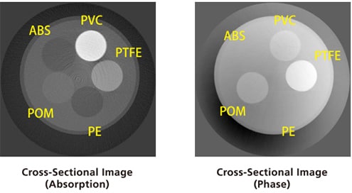
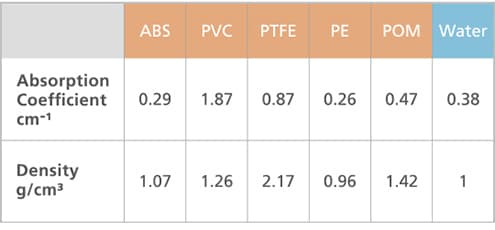
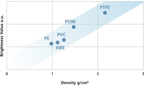
Water and Multiple Types of Resins
The contrast differs for the absorption image and the phase image. As shown in the graphs below, it is evident that the brightness value for the absorption image is proportional to the absorption coefficient, and the brightness value for the phase image is proportional to the density.

Applications (Phase Images)
Mouse (Neonate)
This is a sample scan of a mouse (neonate). The internal organs can be observed without a contrast medium by, just using formalin fixation.
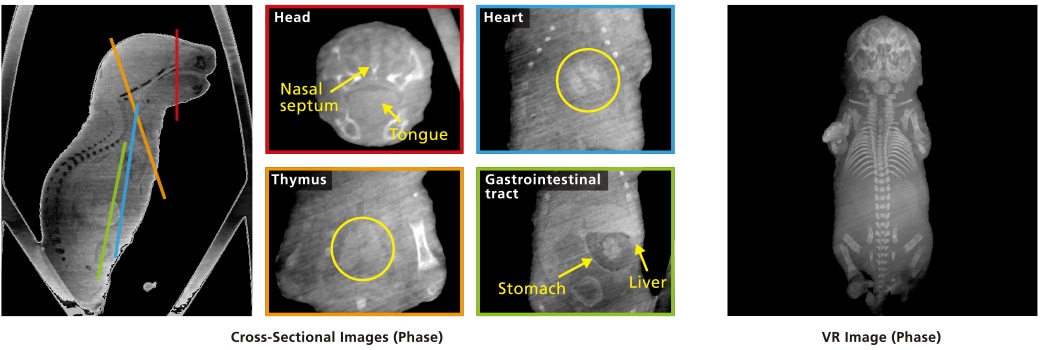
A single scan with Xctal 5000 provides the internal feature of soft tissues such as tongue, heart, thymus, liver, stomach, as well as the clear image of hard tissues equivalent to that scanned by a conventional micro CT system.
The sample remains intact without using contrast enhancing media, enabling users to carry out further histological analyses immediately.
(Samples and comments provided by: Professor Sachiko Iseki and Shigeru Okuhara, Section of Molecular Craniofacial Embryology and Oral Histology, Tokyo Medical and Dental University)
The affiliation of the providers of the samples and comments is as of November 11, 2022.
Insects and Fish
These are sample scans of a robust cicada and twospot hogfish.
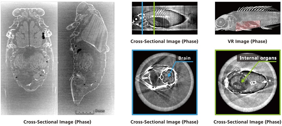
Insects have conventionally been too small for high-resolution CT scans to be performed. In these cross-sectional images, however, the flight muscle bundles running across the thorax of a female robust cicada and individual eggs packed into the abdomen are clearly visible.
(Samples and comments provided by: Dr. Shuhei Nomura, Division of Terrestrial Invertebrates, National Museum of Nature and Science)
This system is capable of observing the viscera and brains of small vertebrates, such as fishes, morphologically. As a result, it is expected to advance progress in the difficult surveys of soft parts. The new observative method does not require staining, which is important for maintaining precious biological specimens and materials.
(Samples and comments provided by: Dr. Gento Shinohara, Division of Vertebrates, National Museum of Nature and Science)
The affiliation of the providers of the samples and comments is as of July 27, 2022.
Anyone Can Easily Perform New Observations
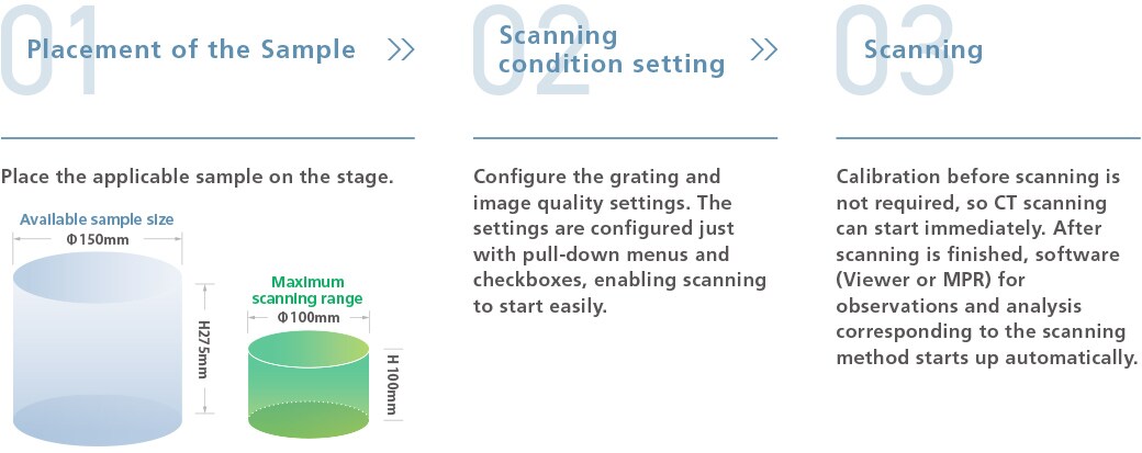
Three-Step Scan
Standard Software
-
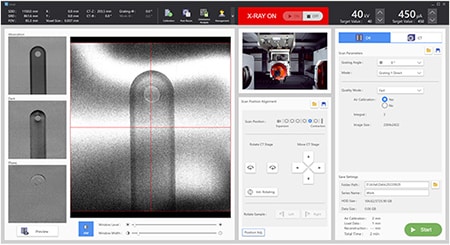
Xctal
Functions include system control, fluoroscopy, CT scan, and reconstitution.
Control Software -
-
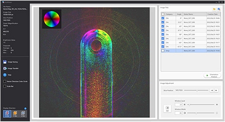
XctalViewer
This software displays the X-ray 2D projection images and cross-sectional images from scans with Xctal 5000.
Viewer Software
In addition to displaying images, it can output vector direction color maps created from multiple dark-field images. -
-
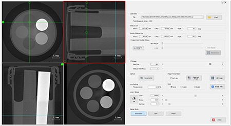
MPR(Xctal)
This software displays cross-sectional images obtained with the Xctal 5000. Four cross-sectional images at any angle can be displayed simultaneously. -