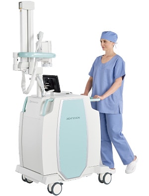
Near-Infrared Fluorescence Imaging System - LIGHTVISION
![]()
![]()
*Transfers to the Virtual Exhibition site.
High Definition Near-Infrared Fluorescence Imaging System
This page introduces a surgery support system for medical professionals and includes clinical images during surgery.
Please contact Shimadzu local representative for product availability.

ICG Fluorescence using Near-infrared light
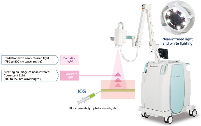
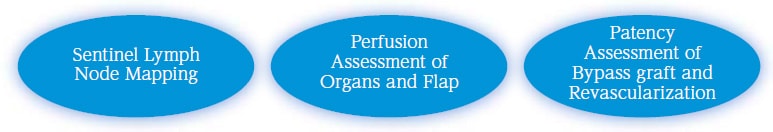
The near-infrared fluorescence imaging system irradiates near-infrared excitation light on an ICG (indocyanine green) contrast medium injected into the blood vessels, lymph ducts, or other areas. It is a system that allows the observation of blood flow and lymph flow under the tissue surface by imaging the near-infrared fluorescence. This method, called ICG fluorescence imaging, enables the simple and real time identification of lymphatic vessels and the confirmation of blood flow during surgery. It has been confirmed to be useful for the quick localization of sentinel lymph nodes and, tissue and organ perfusion.
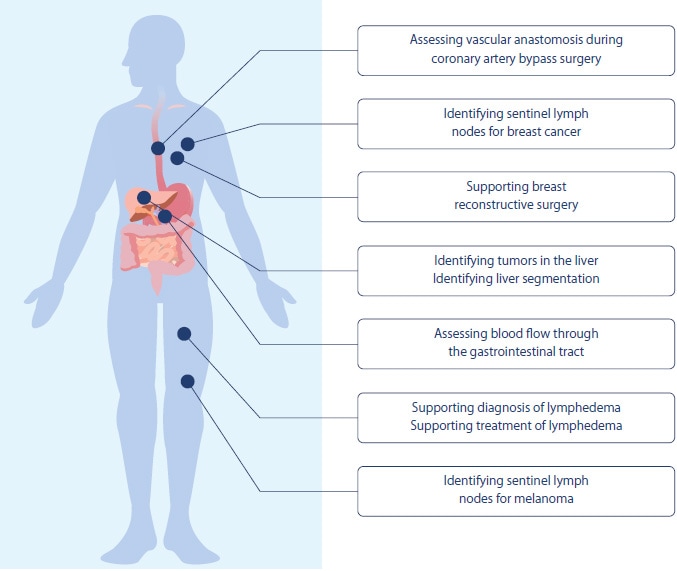
Expanding Clinical Applications
Shimadzu’s LIGHTVISION system provides unprecedented clinical value by easily visualizing lymph and blood flow during surgery.
Equipped with high definition sensors, the LIGHTVISION system displays detailed high definition images, which are especially useful for procedures requiring confirmation of blood flow through small blood vessels, such as in surgical flaps. Due to the large field of view, an image of a broad area, such as the abdomen, can be displayed on one screen, providing a quick overview when assessing blood flow through blood vessels in an abdominal flap or the gastric tube. Furthermore, the colors displayed in fluorescence images can be changed, which is especially helpful for identifying sentinel lymph nodes. Consequently, clinical applications of LIGHTVISION are expanding into a wide variety of surgical procedures.
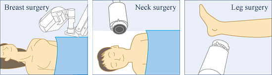
Plastic and Reconstructive Surgery
【Application】Assessing blood flow through a surgical flap
The 10x zoom function provides a wide viewing angle, capturing a large field of view, including an entire large abdominal flap displayed on one screen, which is helpful for judging flap perfusion. An enlarged view can be used for high definition visualization of blood vessels to assess patency after anastomosis.
Breast Reconstruction Assessment of Flap Perfusion
Breast Reconstruction Assessment of Blood Vessel Patency
【Application】Diagnosis and treatment of lymphedema
LIGHTVISION can support the determination of the progression of lymphedema by visualizing the pattern of travel through lymphatic vessels. During lymphovenous anastomosis(LVA) procedures, both near-infrared and visible images can be referenced to easily mark the lymphatic vessels.
Marking Lymphatic Vessels During LVA
Dermatologic Surgery
【Application】Identifying sentinel lymph nodes for melanoma
Because LIGHTVISION is able to clearly display a large area on a single monitor screen, it can be used to quickly and easily trace the flow of lymph extending far from the primary cancer lesion.
Melanoma Sentinel Lymph Node Biopsy
Breast Surgery
【Application】Identifying Application sentinel lymph nodes
LIGHTVISION helps identify sentinel lymph nodes accurately by clearly visualizing lymph flow over a broad area, extending from the primary lesion to the axilla. After incision, the zoom function can be used to display a magnified view of the incised area, so that lymphatic vessel can be visually identified during excision. Since it does not use any radioactive substance, no radiation exposure is involved.
Breast Cancer Sentinel lymph Node Biopsy
Image provided by:Kochi Medical School Hospital Breast Center, Japan
Cardiovascular Surgery
【Application】Assessing the patency of CABG
LIGHTVISION can be used for intraoperative assessment of the coronary artery bypass graft patency and the perfusion of blood through the myocardium during surgery.
Coronary Artery Bypass Grafting (CABG) Assessing Blood Vessel Patency and Myocardial Perfusion
Image provided by:Kochi Medical School Hospital, Japan
Hepato-Pancreato-Biliary Surgery
【Application】Identifying tumors in the liver
Indocyanine green (ICG), used to examine liver function before surgery, remains unmetabolized in tumors and other areas with abnormal liver cells. This characteristic can be used to support more reliable and minimally invasive surgery
by identifying tumors, determining the margins for excision.
Liver Tumor Excising a Tumorous Area from a Liver
【Application】Identifying the liver segmentation
By administering the ICG via a branch of portal vein, the affected region of the liver can be clearly visualized and the demarcation line for excision can be decided accurately.
Liver Tumor Hepatic Segmental Resection
Upper Gastrointestinal Surgery
【Application】Assessing blood flow through the gastric tube
Blood perfusion through tissue can be visualized with clear image quality. By visualizing vascularity during surgery with clear image quality, it visually supports deciding areas for anastomosis.
Esophagus Cancer, Assessing Perfusion of Gastric Tube Tissue
Image provided by:Kochi Medical School Department of Surgery ,Japan
Colorectal Surgery
【Application】Assessing tissue perfusion during colon cancer surgery
Colorectal Surgery, Assessing tissue perfusion during colon cancer surgery
Accurate Surgical Navigation with High Definition Images
High Definition Image Quality
Equipped with high definition sensors, the system can display detailed high definition images. In addition to clearly confirming blood vessels and lymph ducts emitting fluorescent light, the high-resolution images can also be used to confirm the positional relationship between surrounding tissue and surgical tools during surgery. Given the accuracy and speed demands of the surgical environment, this system assists in making more accurate decisions more rapidly.
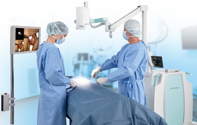
Display Images Clearly on a Large Monitor
It is possible to display the images on a large remotely located monitor to allow information sharing with all the surgical staff.
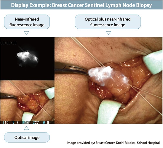
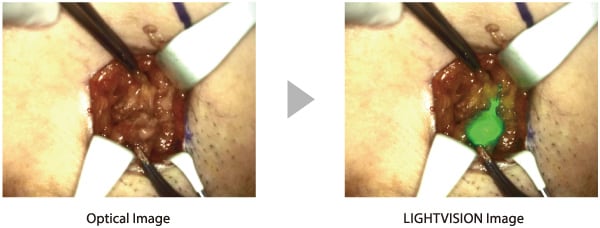
Simultaneous Three-Image Display Provides Clear Understanding at a Glance
The system can display three images, an Optical image, a near-infrared fluorescence image, and a combined visible plus near-infrared fluorescence image, at the same time. This view enables confirmation of the area with ICG illumination by comparing it to the surrounding tissue, which is especially effective for determining the ablation range.
This enables a large amount of information to be referenced and confirmed at a glance during surgery, which can help shorten procedure times.
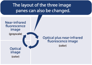
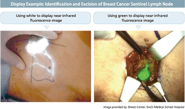
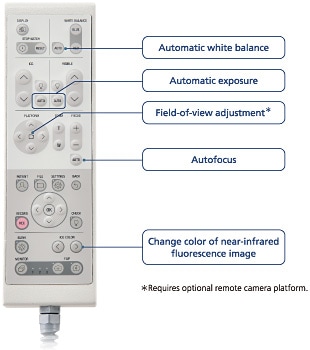
Colors Can Be Changed to Improve the Visibility of Blood Vessel or Lymphatic Vessel
To improve the visualization of blood vessels or lymph ducts, the color for the near-infrared fluorescence image can be changed to a white, green or blue color that is easier to distinguish from the surrounding tissues or light reflection of blood vessels or body fluids.
An easy to use button is used to change the colour, so that the optimum color can be selected according to the condition of the surgical site.
Imaging is Possible in a Bright Room
Images can be acquired with the operating room lighting left ON, which means images can be compared to the affected area.
- *Surgical lights must be switched OFF.
Excellent Operability Allows for Concentration on Surgery
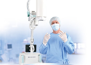
Hands-Free Imaging
The self-supported extendable arm allows hands-free operation. In addition to reducing the burden on surgical personnel, it can also display images without blur due to hand movement.
Wide Camera Movement Range
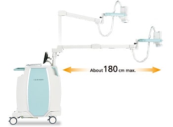
Freely Adjustable Camera Positioning
With a fully balanced arm, you can quickly position the camera height and angle as you wish. Images can be acquired via the optimal camera position depending on the type of surgery or condition of the affected area.
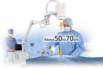
Ensures Working Distance Required for Surgical Work
Due to the bright light source and high sensitivity image sensor, a working distance (distance between the surgical field and camera) of about 50 to 70 cm can be maintained during imaging. Consequently, imaging can be performed at the same time as working in the surgical field.
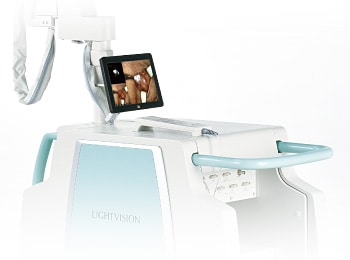
Images Can Be Confirmed on a Small Monitor on the Main Unit
The operator can check the system operating situation and adjust image quality with a small monitor on the main unit.
Camera Adjustments by Remote Control
Camera adjustment by remote control is possible. The remote control includes three automatic adjustment functions :
exposure, focus, and white balance, which quickly optimize the image display.
Main Unit Is Easy to Move and Reposition
The main unit is designed so that it can be moved reliably between operating rooms. Handles are provided on the front and back, so that the unit can be easily repositioned within the operating room.
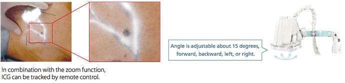
Remote Adjustable Field-of-View Camera Platform(optional)
An optional camera platform is available for adjusting the camera angle by remote control.
It can be operated from a location away from the main unit to adjust the camera field of view, so that the area of interest is displayed.
The product design may be partially changed without notice.
No FDA(510k) or CE clearance. Please contact us to confirm the availability of this product in your country.
References
Yoshimatsu H. et al. Application of intraoperative indocyanine green angiography for detecting flap congestion in the use of free deep inferior epigastric perforator flaps for breast reconstruction
Microsurgery 2021
Karakawa R. et al. Supermicrosurgical Suture-Stent Technique for a Lymphaticovenular Bypass
Clinical Medicine 2021
Yamamoto M. et al. The impact of the quantitative assessment procedure for coronary artery bypass graft evaluations using high-resolution near-infrared fluorescence angiography
Surgery Today 2021
Kitagawa H. et al. Correlation between indocyanine green visualization time in the gastric tube and postoperative endoscopic assessment of the anastomosis after esophageal surgery
Surgery Today 2020
Kusada T. et al. Different indocyanine green fluorescence patterns of two skin metastases of hypopharyngeal squamous carcinoma: A case report
Photodiagnosis and Photodynamic Therapy 2021
Kusada T. et al. Indocyanine green fluorescence angiography for detection of cutaneous angiosarcoma of the scalp: A case report
Photodiagnosis and Photodynamic Therapy 2020
Seki Y. et al. Real-time Indocyanine Green Videolymphography Navigation for Lymphaticovenular Anastomosis
Plastic and Reconstructive Surgery-Global Open 2019
Teramoto Y. et al. Intraoperative use of LIGHTVISION: a novel fluorescence navigation system using indocyanine green for sentinel lymph node biopsy in skin cancer patients.
European Journal of Dermatology 2018
Miyashita S. et al. Hepatectomy Using a Novel Cart-Based Indocyanine Green Fluorescence Imaging System
Surgery Today
Tsukuura R. et al. Novel hands‐free near‐infrared fluorescence navigation and simultaneous combined imaging for elevation of vascularized lymph node flap
Journal of Surgical Oncology

Saitama Jikei Hospital/Saitama Hand & Microsurgery Institute
Lymphedema Clinic, Department of Breast Center, Kameda Medical Center
Lymphedema Clinic, Kameda Kyobashi Clinic
Breast Center, Kochi Medical School Hospital